7FAB

CRYSTAL STRUCTURE OF HUMAN IMMUNOGLOBULIN FRAGMENT FAB NEW REFINED AT 2.0 ANGSTROMS RESOLUTION


Experimental Data Snapshot

  • Method: X-RAY DIFFRACTION
  • Resolution: 2.00 Å
  • R-Value Work: 0.169 
  • R-Value Observed: 0.169 

wwPDB Validation   3D Report Full Report


This is version 1.3 of the entry. See complete history


Literature

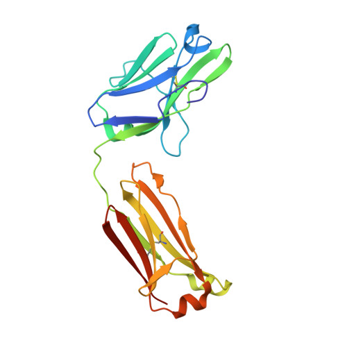
Crystal structure of human immunoglobulin fragment Fab New refined at 2.0 A resolution.

Saul, F.A.Poljak, R.J.

(1992) Proteins 14: 363-371

  • DOI: https://doi.org/10.1002/prot.340140305
  • Primary Citation of Related Structures:  
    7FAB

  • PubMed Abstract: 

    The three-dimensional structure of the human immunoglobulin fragment Fab New (IgG1, lambda) has been refined to a crystallographic R-factor of 16.9% to 2 A resolution. Rms deviations of the final model from ideal geometry are 0.014 A for bond distances and 3.03 degrees for bond angles. Refinement was based on a new X-ray data set including 28,301 reflections with F > 2.5 sigma(F) from 6.0 to 2.0 A resolution. The starting model for the refinement procedure reported here is from the Brookhaven Protein Data Bank entry 3FAB (rev. 1981). Differences between the initial and final models include modified polypeptide-chain folding in the third complementarity-determining region (CDR3) and the third framework region (FR3) of VH and in some exposed loops of CL and CH1. Amino acid sequence changes were determined at a number of positions by inspection of difference electron density maps. The incorporation of amino acid sequence changes results in an improved VH framework model for the "humanization" of monoclonal antibodies.


  • Organizational Affiliation

    Département d'Immunologie, Institut Pasteur, Paris, France.


Macromolecules
Find similar proteins by:  (by identity cutoff)  |  3D Structure
Entity ID: 1
MoleculeChains Sequence LengthOrganismDetailsImage
IGG1-LAMBDA NEW FAB (LIGHT CHAIN)A [auth L]208Homo sapiensMutation(s): 0 
UniProt & NIH Common Fund Data Resources
Find proteins for P0DOY2 (Homo sapiens)
Explore P0DOY2 
Go to UniProtKB:  P0DOY2
GTEx:  ENSG00000211677 
Entity Groups  
Sequence Clusters30% Identity50% Identity70% Identity90% Identity95% Identity100% Identity
UniProt GroupP0DOY2
Sequence Annotations
Expand
  • Reference Sequence
Find similar proteins by:  (by identity cutoff)  |  3D Structure
Entity ID: 2
MoleculeChains Sequence LengthOrganismDetailsImage
IGG1-LAMBDA NEW FAB (HEAVY CHAIN)B [auth H]217Homo sapiensMutation(s): 0 
UniProt & NIH Common Fund Data Resources
Find proteins for P01825 (Homo sapiens)
Explore P01825 
Go to UniProtKB:  P01825
GTEx:  ENSG00000224373 
Entity Groups  
Sequence Clusters30% Identity50% Identity70% Identity90% Identity95% Identity100% Identity
UniProt GroupP01825
Sequence Annotations
Expand
  • Reference Sequence
Experimental Data & Validation

Experimental Data

  • Method: X-RAY DIFFRACTION
  • Resolution: 2.00 Å
  • R-Value Work: 0.169 
  • R-Value Observed: 0.169 
  • Space Group: C 1 2 1
Unit Cell:
Length ( Å )Angle ( ˚ )
a = 111.43α = 90
b = 56.68β = 116.46
c = 90.3γ = 90
Software Package:
Software NamePurpose
X-PLORmodel building
X-PLORrefinement
X-PLORphasing

Structure Validation

View Full Validation Report


Entry History 

Deposition Data

Revision History  (Full details and data files)

  • Version 1.0: 1994-01-31
    Type: Initial release
  • Version 1.1: 2008-03-25
    Changes: Version format compliance
  • Version 1.2: 2011-07-13
    Changes: Version format compliance
  • Version 1.3: 2013-07-24
    Changes: Database references, Structure summary