7F1E

Structure of METTL6 bound with SAM


Experimental Data Snapshot

  • Method: X-RAY DIFFRACTION
  • Resolution: 2.59 Å
  • R-Value Free: 0.227 
  • R-Value Work: 0.191 
  • R-Value Observed: 0.193 

wwPDB Validation   3D Report Full Report


Ligand Structure Quality Assessment 


This is version 1.1 of the entry. See complete history


Literature

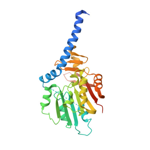
Structural basis for METTL6-mediated m3C RNA methylation.

Li, S.Zhou, H.Liao, S.Wang, X.Zhu, Z.Zhang, J.Xu, C.

(2021) Biochem Biophys Res Commun 589: 159-164

  • DOI: https://doi.org/10.1016/j.bbrc.2021.12.013
  • Primary Citation of Related Structures:  
    7F1E

  • PubMed Abstract: 

    RNA modifications play important roles in mediating the biological functions of RNAs. 3-methylcytidine (m3C), albeit less abundant, is found to exist extensively in tRNAs, rRNAs and mRNAs. Human METTL6 is a m 3 C methyltransferase for tRNAs, including tRNA SER(UGA) . We solved the structure of human METTL6 in the presence of S-adenosyl-L-methionine and found by enzyme assay that recombinant human METTL6 is active towards tRNA SER(UGA) . Structural analysis indicated the detailed interactions between S-adenosyl-L-methionine and METTL6, and suggested potential tRNA binding region on the surface of METTL6. The structural research, complemented by biochemistry enzyme assay, will definitely shed light on the design of potent inhibitors for METTL6 in near future.


  • Organizational Affiliation

    MOE Key Laboratory for Membraneless Organelles & Cellular Dynamics, Hefei National Laboratory for Physical Sciences at the Microscale, School of Life Sciences, Division of Life Sciences and Medicine, University of Science and Technology of China, 230027, Hefei, PR China.


Macromolecules
Find similar proteins by:  (by identity cutoff)  |  3D Structure
Entity ID: 1
MoleculeChains Sequence LengthOrganismDetailsImage
tRNA N(3)-methylcytidine methyltransferase METTL6
A, B
286Homo sapiensMutation(s): 0 
Gene Names: METTL6
EC: 2.1.1
UniProt & NIH Common Fund Data Resources
Find proteins for Q8TCB7 (Homo sapiens)
Explore Q8TCB7 
Go to UniProtKB:  Q8TCB7
PHAROS:  Q8TCB7
GTEx:  ENSG00000206562 
Entity Groups  
Sequence Clusters30% Identity50% Identity70% Identity90% Identity95% Identity100% Identity
UniProt GroupQ8TCB7
Sequence Annotations
Expand
  • Reference Sequence
Experimental Data & Validation

Experimental Data

  • Method: X-RAY DIFFRACTION
  • Resolution: 2.59 Å
  • R-Value Free: 0.227 
  • R-Value Work: 0.191 
  • R-Value Observed: 0.193 
  • Space Group: P 43
Unit Cell:
Length ( Å )Angle ( ˚ )
a = 83.457α = 90
b = 83.457β = 90
c = 129.686γ = 90
Software Package:
Software NamePurpose
PHENIXrefinement
PDB_EXTRACTdata extraction
HKL-2000data reduction
HKL-2000data scaling
PHASERphasing

Structure Validation

View Full Validation Report


Ligand Structure Quality Assessment 


Entry History & Funding Information

Deposition Data


Funding OrganizationLocationGrant Number
National Natural Science Foundation of China (NSFC)China31770806

Revision History  (Full details and data files)

  • Version 1.0: 2022-01-12
    Type: Initial release
  • Version 1.1: 2023-11-29
    Changes: Data collection, Refinement description