7DCM

Crystal structure of CITX


Experimental Data Snapshot

  • Method: X-RAY DIFFRACTION
  • Resolution: 2.50 Å
  • R-Value Free: 0.257 
  • R-Value Work: 0.171 
  • R-Value Observed: 0.179 

wwPDB Validation   3D Report Full Report


This is version 1.1 of the entry. See complete history


Literature

Co-evolution-based prediction of metal-binding sites in proteomes by machine learning.

Cheng, Y.Wang, H.Xu, H.Liu, Y.Ma, B.Chen, X.Zeng, X.Wang, X.Wang, B.Shiau, C.Ovchinnikov, S.Su, X.D.Wang, C.

(2023) Nat Chem Biol 19: 548-555

  • DOI: https://doi.org/10.1038/s41589-022-01223-z
  • Primary Citation of Related Structures:  
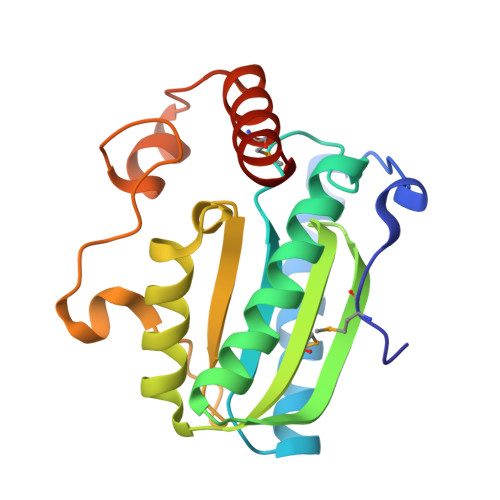
    7DCM, 7DCN

  • PubMed Abstract: 

    Metal ions have various important biological roles in proteins, including structural maintenance, molecular recognition and catalysis. Previous methods of predicting metal-binding sites in proteomes were based on either sequence or structural motifs. Here we developed a co-evolution-based pipeline named 'MetalNet' to systematically predict metal-binding sites in proteomes. We applied MetalNet to proteomes of four representative prokaryotic species and predicted 4,849 potential metalloproteins, which substantially expands the currently annotated metalloproteomes. We biochemically and structurally validated previously unannotated metal-binding sites in several proteins, including apo-citrate lyase phosphoribosyl-dephospho-CoA transferase citX, an Escherichia coli enzyme lacking structural or sequence homology to any known metalloprotein (Protein Data Bank (PDB) codes: 7DCM and 7DCN ). MetalNet also successfully recapitulated all known zinc-binding sites from the human spliceosome complex. The pipeline of MetalNet provides a unique and enabling tool for interrogating the hidden metalloproteome and studying metal biology.


  • Organizational Affiliation

    Synthetic and Functional Biomolecules Center, Beijing National Laboratory for Molecular Sciences, Key Laboratory of Bioorganic Chemistry and Molecular Engineering of Ministry of Education, Peking University, Beijing, China.


Macromolecules
Find similar proteins by:  (by identity cutoff)  |  3D Structure
Entity ID: 1
MoleculeChains Sequence LengthOrganismDetailsImage
Probable apo-citrate lyase phosphoribosyl-dephospho-CoA transferase186Escherichia coliMutation(s): 0 
EC: 2.7.7.61
UniProt
Find proteins for P0A6G5 (Escherichia coli (strain K12))
Explore P0A6G5 
Go to UniProtKB:  P0A6G5
Entity Groups  
Sequence Clusters30% Identity50% Identity70% Identity90% Identity95% Identity100% Identity
UniProt GroupP0A6G5
Sequence Annotations
Expand
  • Reference Sequence
Small Molecules
Ligands 1 Unique
IDChains Name / Formula / InChI Key2D Diagram3D Interactions
ZN (Subject of Investigation/LOI)
Query on ZN

Download Ideal Coordinates CCD File 
B [auth A]ZINC ION
Zn
PTFCDOFLOPIGGS-UHFFFAOYSA-N
Modified Residues  1 Unique
IDChains TypeFormula2D DiagramParent
MSE
Query on MSE
A
L-PEPTIDE LINKINGC5 H11 N O2 SeMET
Experimental Data & Validation

Experimental Data

  • Method: X-RAY DIFFRACTION
  • Resolution: 2.50 Å
  • R-Value Free: 0.257 
  • R-Value Work: 0.171 
  • R-Value Observed: 0.179 
  • Space Group: P 1 21 1
Unit Cell:
Length ( Å )Angle ( ˚ )
a = 43.017α = 90
b = 36.784β = 92.13
c = 48.265γ = 90
Software Package:
Software NamePurpose
HKL-2000data reduction
HKL-2000data scaling
PHENIXrefinement
PDB_EXTRACTdata extraction
AutoSolphasing

Structure Validation

View Full Validation Report


Entry History 

Deposition Data

Revision History  (Full details and data files)

  • Version 1.0: 2021-11-03
    Type: Initial release
  • Version 1.1: 2023-05-17
    Changes: Database references