7B9K

Cryo-EM structure of the dihydrolipoyl transacetylase cubic core of the E. coli pyruvate dehydrogenase complex including lipoyl domains


Experimental Data Snapshot

  • Method: ELECTRON MICROSCOPY
  • Resolution: 3.16 Å
  • Aggregation State: PARTICLE 
  • Reconstruction Method: SINGLE PARTICLE 

wwPDB Validation   3D Report Full Report


This is version 1.1 of the entry. See complete history


Literature

Structure of the native pyruvate dehydrogenase complex reveals the mechanism of substrate insertion.

Skerlova, J.Berndtsson, J.Nolte, H.Ott, M.Stenmark, P.

(2021) Nat Commun 12: 5277-5277

  • DOI: https://doi.org/10.1038/s41467-021-25570-y
  • Primary Citation of Related Structures:  
    7B9K

  • PubMed Abstract: 

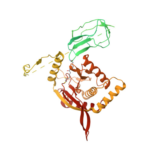
    The pyruvate dehydrogenase complex (PDHc) links glycolysis to the citric acid cycle by converting pyruvate into acetyl-coenzyme A. PDHc encompasses three enzymatically active subunits, namely pyruvate dehydrogenase, dihydrolipoyl transacetylase, and dihydrolipoyl dehydrogenase. Dihydrolipoyl transacetylase is a multidomain protein comprising a varying number of lipoyl domains, a peripheral subunit-binding domain, and a catalytic domain. It forms the structural core of the complex, provides binding sites for the other enzymes, and shuffles reaction intermediates between the active sites through covalently bound lipoyl domains. The molecular mechanism by which this shuttling occurs has remained elusive. Here, we report a cryo-EM reconstruction of the native E. coli dihydrolipoyl transacetylase core in a resting state. This structure provides molecular details of the assembly of the core and reveals how the lipoyl domains interact with the core at the active site.


  • Organizational Affiliation

    Department of Biochemistry and Biophysics, Stockholm University, Stockholm, Sweden.


Macromolecules
Find similar proteins by:  (by identity cutoff)  |  3D Structure
Entity ID: 1
MoleculeChains Sequence LengthOrganismDetailsImage
Dihydrolipoyllysine-residue acetyltransferase component of pyruvate dehydrogenase complex
A, B, C, D, E
A, B, C, D, E, F, G, H, I, J, K, L, M, N, O, P, Q, R, S, T, U, V, W, X
630Escherichia coli K-12Mutation(s): 0 
EC: 2.3.1.12
UniProt
Find proteins for P06959 (Escherichia coli (strain K12))
Explore P06959 
Go to UniProtKB:  P06959
Entity Groups  
Sequence Clusters30% Identity50% Identity70% Identity90% Identity95% Identity100% Identity
UniProt GroupP06959
Sequence Annotations
Expand
  • Reference Sequence
Small Molecules
Modified Residues  1 Unique
IDChains TypeFormula2D DiagramParent
LA2
Query on LA2
A, B, C, D, E
A, B, C, D, E, F, G, H, I, J, K, L, M, N, O, P, Q, R, S, T, U, V, W, X
L-PEPTIDE LINKINGC14 H28 N2 O3 S2LYS
Experimental Data & Validation

Experimental Data

  • Method: ELECTRON MICROSCOPY
  • Resolution: 3.16 Å
  • Aggregation State: PARTICLE 
  • Reconstruction Method: SINGLE PARTICLE 
EM Software:
TaskSoftware PackageVersion
RECONSTRUCTIONcryoSPARC2.15
MODEL REFINEMENTPHENIX1.16
MODEL REFINEMENTREFMAC5.8.0232

Structure Validation

View Full Validation Report


Entry History & Funding Information

Deposition Data


Funding OrganizationLocationGrant Number
Swedish Research CouncilSweden2018-03406
European Regional Development FundEuropean Union"IOCB Mobility" CZ.02.2.69/0.0/0.0/16_027/0008477

Revision History  (Full details and data files)

  • Version 1.0: 2021-08-11
    Type: Initial release
  • Version 1.1: 2021-10-06
    Changes: Data collection, Database references