6Z4X

Structure of the CAK complex form Chaetomium thermophilum bound to ATP-gamma-S


Experimental Data Snapshot

  • Method: X-RAY DIFFRACTION
  • Resolution: 2.98 Å
  • R-Value Free: 0.246 
  • R-Value Work: 0.203 
  • R-Value Observed: 0.205 

wwPDB Validation   3D Report Full Report


Ligand Structure Quality Assessment 


This is version 1.1 of the entry. See complete history


Literature

Structural basis for CDK7 activation by MAT1 and Cyclin H.

Peissert, S.Schlosser, A.Kendel, R.Kuper, J.Kisker, C.

(2020) Proc Natl Acad Sci U S A 117: 26739-26748

  • DOI: https://doi.org/10.1073/pnas.2010885117
  • Primary Citation of Related Structures:  
    6Z3U, 6Z4X

  • PubMed Abstract: 

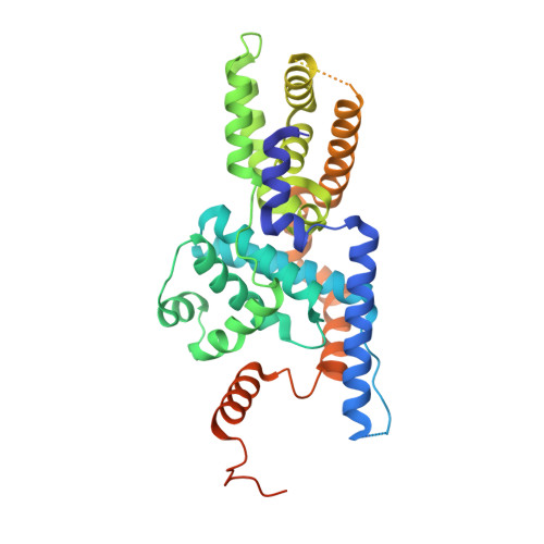
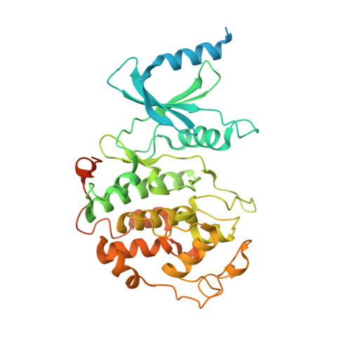
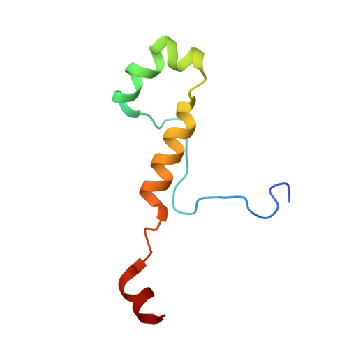
    Cyclin-dependent kinase 7 (CDK7), Cyclin H, and the RING-finger protein MAT1 form the heterotrimeric CDK-activating kinase (CAK) complex which is vital for transcription and cell-cycle control. When associated with the general transcription factor II H (TFIIH) it activates RNA polymerase II by hyperphosphorylation of its C-terminal domain (CTD). In the absence of TFIIH the trimeric complex phosphorylates the T-loop of CDKs that control cell-cycle progression. CAK holds a special position among the CDK branch due to this dual activity and the dependence on two proteins for activation. We solved the structure of the CAK complex from the model organism Chaetomium thermophilum at 2.6-Å resolution. Our structure reveals an intricate network of interactions between CDK7 and its two binding partners MAT1 and Cyclin H, providing a structural basis for the mechanism of CDK7 activation and CAK activity regulation. In vitro activity measurements and functional mutagenesis show that CDK7 activation can occur independent of T-loop phosphorylation and is thus exclusively MAT1-dependent by positioning the CDK7 T-loop in its active conformation.


  • Organizational Affiliation

    Rudolf Virchow Center for Integrative and Translational Bioimaging, Institute for Structural Biology, University of Würzburg, 97080 Würzburg, Germany.


Macromolecules
Find similar proteins by:  (by identity cutoff)  |  3D Structure
Entity ID: 1
MoleculeChains Sequence LengthOrganismDetailsImage
CYCLIN domain-containing protein
A, D
425Thermochaetoides thermophilaMutation(s): 0 
UniProt
Find proteins for G0SH78 (Chaetomium thermophilum (strain DSM 1495 / CBS 144.50 / IMI 039719))
Explore G0SH78 
Go to UniProtKB:  G0SH78
Entity Groups  
Sequence Clusters30% Identity50% Identity70% Identity90% Identity95% Identity100% Identity
UniProt GroupG0SH78
Sequence Annotations
Expand
  • Reference Sequence
Find similar proteins by:  (by identity cutoff)  |  3D Structure
Entity ID: 2
MoleculeChains Sequence LengthOrganismDetailsImage
Protein kinase domain-containing protein
B, E
437Thermochaetoides thermophilaMutation(s): 0 
UniProt
Find proteins for G0SFC6 (Chaetomium thermophilum (strain DSM 1495 / CBS 144.50 / IMI 039719))
Explore G0SFC6 
Go to UniProtKB:  G0SFC6
Entity Groups  
Sequence Clusters30% Identity50% Identity70% Identity90% Identity95% Identity100% Identity
UniProt GroupG0SFC6
Sequence Annotations
Expand
  • Reference Sequence
Find similar proteins by:  (by identity cutoff)  |  3D Structure
Entity ID: 3
MoleculeChains Sequence LengthOrganismDetailsImage
RING-type domain-containing protein
C, F
69Thermochaetoides thermophilaMutation(s): 0 
UniProt
Find proteins for G0SF48 (Chaetomium thermophilum (strain DSM 1495 / CBS 144.50 / IMI 039719))
Explore G0SF48 
Go to UniProtKB:  G0SF48
Entity Groups  
Sequence Clusters30% Identity50% Identity70% Identity90% Identity95% Identity100% Identity
UniProt GroupG0SF48
Sequence Annotations
Expand
  • Reference Sequence
Experimental Data & Validation

Experimental Data

  • Method: X-RAY DIFFRACTION
  • Resolution: 2.98 Å
  • R-Value Free: 0.246 
  • R-Value Work: 0.203 
  • R-Value Observed: 0.205 
  • Space Group: P 1 21 1
Unit Cell:
Length ( Å )Angle ( ˚ )
a = 89.97α = 90
b = 84.903β = 96.197
c = 154.477γ = 90
Software Package:
Software NamePurpose
BUSTERrefinement
PHENIXrefinement
XDSdata reduction
STARANISOdata scaling
PHASERphasing

Structure Validation

View Full Validation Report


Ligand Structure Quality Assessment 


Entry History 

Deposition Data

Revision History  (Full details and data files)

  • Version 1.0: 2020-12-09
    Type: Initial release
  • Version 1.1: 2024-01-24
    Changes: Data collection, Database references, Refinement description