6YXA

Structure of the bifunctional Rel enzyme from B. subtilis


Experimental Data Snapshot

  • Method: X-RAY DIFFRACTION
  • Resolution: 3.95 Å
  • R-Value Free: 0.284 
  • R-Value Work: 0.264 
  • R-Value Observed: 0.266 

wwPDB Validation   3D Report Full Report


This is version 1.2 of the entry. See complete history


Literature

Structural Basis for Regulation of the Opposing (p)ppGpp Synthetase and Hydrolase within the Stringent Response Orchestrator Rel.

Pausch, P.Abdelshahid, M.Steinchen, W.Schafer, H.Gratani, F.L.Freibert, S.A.Wolz, C.Turgay, K.Wilson, D.N.Bange, G.

(2020) Cell Rep 32: 108157-108157

  • DOI: https://doi.org/10.1016/j.celrep.2020.108157
  • Primary Citation of Related Structures:  
    6HTQ, 6YXA

  • PubMed Abstract: 

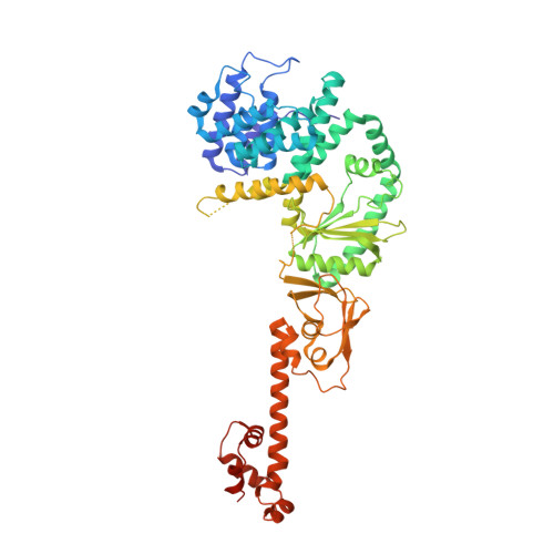
    The stringent response enables metabolic adaptation of bacteria under stress conditions and is governed by RelA/SpoT Homolog (RSH)-type enzymes. Long RSH-type enzymes encompass an N-terminal domain (NTD) harboring the second messenger nucleotide (p)ppGpp hydrolase and synthetase activity and a stress-perceiving and regulatory C-terminal domain (CTD). CTD-mediated binding of Rel to stalled ribosomes boosts (p)ppGpp synthesis. However, how the opposing activities of the NTD are controlled in the absence of stress was poorly understood. Here, we demonstrate on the RSH-type protein Rel that the critical regulative elements reside within the TGS (ThrRS, GTPase, and SpoT) subdomain of the CTD, which associates to and represses the synthetase to concomitantly allow for activation of the hydrolase. Furthermore, we show that Rel forms homodimers, which appear to control the interaction with deacylated-tRNA, but not the enzymatic activity of Rel. Collectively, our study provides a detailed molecular view into the mechanism of stringent response repression in the absence of stress.


  • Organizational Affiliation

    Center for Synthetic Microbiology & Department of Chemistry, Hans-Meerwein-Strasse, C07, Philipps-University Marburg, 35043 Marburg, Germany.


Macromolecules
Find similar proteins by:  (by identity cutoff)  |  3D Structure
Entity ID: 1
MoleculeChains Sequence LengthOrganismDetailsImage
GTP pyrophosphokinase562Bacillus subtilis subsp. subtilis str. 168Mutation(s): 0 
Gene Names: relABSU27600
EC: 2.7.6.5
UniProt
Find proteins for O54408 (Bacillus subtilis (strain 168))
Explore O54408 
Go to UniProtKB:  O54408
Entity Groups  
Sequence Clusters30% Identity50% Identity70% Identity90% Identity95% Identity100% Identity
UniProt GroupO54408
Sequence Annotations
Expand
  • Reference Sequence
Small Molecules
Ligands 1 Unique
IDChains Name / Formula / InChI Key2D Diagram3D Interactions
MN
Query on MN

Download Ideal Coordinates CCD File 
B [auth A]MANGANESE (II) ION
Mn
WAEMQWOKJMHJLA-UHFFFAOYSA-N
Experimental Data & Validation

Experimental Data

  • Method: X-RAY DIFFRACTION
  • Resolution: 3.95 Å
  • R-Value Free: 0.284 
  • R-Value Work: 0.264 
  • R-Value Observed: 0.266 
  • Space Group: P 43 21 2
Unit Cell:
Length ( Å )Angle ( ˚ )
a = 130.152α = 90
b = 130.152β = 90
c = 157.621γ = 90
Software Package:
Software NamePurpose
PHENIXrefinement
XDSdata reduction
Aimlessdata scaling
PHASERphasing

Structure Validation

View Full Validation Report


Entry History & Funding Information

Deposition Data


Funding OrganizationLocationGrant Number
German Research Foundation (DFG)GermanySPP1879

Revision History  (Full details and data files)

  • Version 1.0: 2020-09-23
    Type: Initial release
  • Version 1.1: 2020-09-30
    Changes: Database references, Derived calculations
  • Version 1.2: 2024-01-24
    Changes: Data collection, Database references, Refinement description