6Y64

Structure of Sheep Polyomavirus VP1 in complex with 6'-Sialyllactosamine


Experimental Data Snapshot

  • Method: X-RAY DIFFRACTION
  • Resolution: 1.60 Å
  • R-Value Free: 0.182 
  • R-Value Work: 0.152 
  • R-Value Observed: 0.152 

wwPDB Validation   3D Report Full Report


Ligand Structure Quality Assessment 


This is version 2.2 of the entry. See complete history


Literature

Structural Basis and Evolution of Glycan Receptor Specificities within the Polyomavirus Family.

Stroh, L.J.Rustmeier, N.H.Blaum, B.S.Botsch, J.Rossler, P.Wedekink, F.Lipkin, W.I.Mishra, N.Stehle, T.

(2020) mBio 11

  • DOI: https://doi.org/10.1128/mBio.00745-20
  • Primary Citation of Related Structures:  
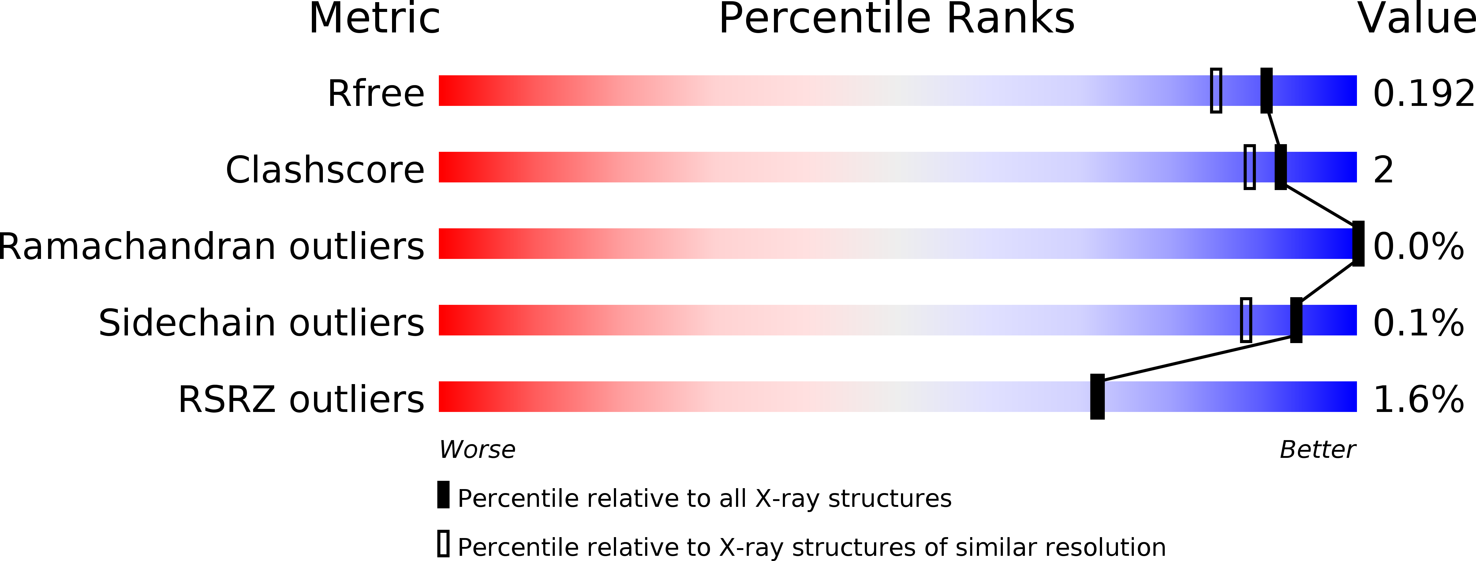
    6Y5X, 6Y5Y, 6Y5Z, 6Y60, 6Y61, 6Y63, 6Y64, 6Y65, 6Y66, 6Y67, 6Y6A, 6Y9I

  • PubMed Abstract: 

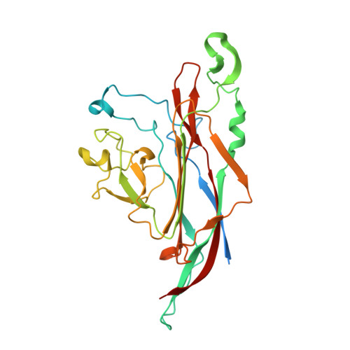
    Asymptomatic infections with polyomaviruses in humans are common, but these small viruses can cause severe diseases in immunocompromised hosts. New Jersey polyomavirus (NJPyV) was identified via a muscle biopsy in an organ transplant recipient with systemic vasculitis, myositis, and retinal blindness, and human polyomavirus 12 (HPyV12) was detected in human liver tissue. The evolutionary origins and potential diseases are not well understood for either virus. In order to define their receptor engagement strategies, we first used nuclear magnetic resonance (NMR) spectroscopy to establish that the major capsid proteins (VP1) of both viruses bind to sialic acid in solution. We then solved crystal structures of NJPyV and HPyV12 VP1 alone and in complex with sialylated glycans. NJPyV employs a novel binding site for a α2,3-linked sialic acid, whereas HPyV12 engages terminal α2,3- or α2,6-linked sialic acids in an exposed site similar to that found in Trichodysplasia spinulosa -associated polyomavirus (TSPyV). Gangliosides or glycoproteins, featuring in mammals usually terminal sialic acids, are therefore receptor candidates for both viruses. Structural analyses show that the sialic acid-binding site of NJPyV is conserved in chimpanzee polyomavirus (ChPyV) and that the sialic acid-binding site of HPyV12 is widely used across the entire polyomavirus family, including mammalian and avian polyomaviruses. A comparison with other polyomavirus-receptor complex structures shows that their capsids have evolved to generate several physically distinct virus-specific receptor-binding sites that can all specifically engage sialylated glycans through a limited number of contacts. Small changes in each site may have enabled host-switching events during the evolution of polyomaviruses. IMPORTANCE Virus attachment to cell surface receptors is critical for productive infection. In this study, we have used a structure-based approach to investigate the cell surface recognition event for New Jersey polyomavirus (NJPyV) and human polyomavirus 12 (HPyV12). These viruses belong to the polyomavirus family, whose members target different tissues and hosts, including mammals, birds, fish, and invertebrates. Polyomaviruses are nonenveloped viruses, and the receptor-binding site is located in their capsid protein VP1. The NJPyV capsid features a novel sialic acid-binding site that is shifted in comparison to other structurally characterized polyomaviruses but shared with a closely related simian virus. In contrast, HPyV12 VP1 engages terminal sialic acids in a manner similar to the human Trichodysplasia spinulosa -associated polyomavirus. Our structure-based phylogenetic analysis highlights that even distantly related avian polyomaviruses possess the same exposed sialic acid-binding site. These findings complement phylogenetic models of host-virus codivergence and may also reflect past host-switching events.


  • Organizational Affiliation

    Interfaculty Institute of Biochemistry, University of Tübingen, Tübingen, Germany.


Macromolecules
Find similar proteins by:  (by identity cutoff)  |  3D Structure
Entity ID: 1
MoleculeChains Sequence LengthOrganismDetailsImage
Capsid protein VP1
A, B, C, D, E
A, B, C, D, E, F, G, H, I, J
293Sheep polyomavirus 1Mutation(s): 0 
UniProt
Find proteins for A0A0E3ZCF3 (Sheep polyomavirus 1)
Explore A0A0E3ZCF3 
Go to UniProtKB:  A0A0E3ZCF3
Entity Groups  
Sequence Clusters30% Identity50% Identity70% Identity90% Identity95% Identity100% Identity
UniProt GroupA0A0E3ZCF3
Sequence Annotations
Expand
  • Reference Sequence
Oligosaccharides

Help

Entity ID: 2
MoleculeChains Length2D Diagram Glycosylation3D Interactions
N-acetyl-alpha-neuraminic acid-(2-6)-beta-D-galactopyranose
K, L, N
2N/A
Glycosylation Resources
GlyTouCan:  G63069TR
GlyCosmos:  G63069TR
GlyGen:  G63069TR
Entity ID: 3
MoleculeChains Length2D Diagram Glycosylation3D Interactions
N-acetyl-alpha-neuraminic acid-(2-6)-beta-D-galactopyranose-(1-4)-2-acetamido-2-deoxy-beta-D-glucopyranose
M
3N/A
Glycosylation Resources
GlyTouCan:  G73578JC
GlyCosmos:  G73578JC
GlyGen:  G73578JC
Small Molecules
Ligands 4 Unique
IDChains Name / Formula / InChI Key2D Diagram3D Interactions
SIA (Subject of Investigation/LOI)
Query on SIA

Download Ideal Coordinates CCD File 
T [auth B],
X [auth C]
N-acetyl-alpha-neuraminic acid
C11 H19 N O9
SQVRNKJHWKZAKO-YRMXFSIDSA-N
GOL
Query on GOL

Download Ideal Coordinates CCD File 
AB [auth I]
BB [auth I]
CA [auth D]
CB [auth I]
DA [auth D]
AB [auth I],
BB [auth I],
CA [auth D],
CB [auth I],
DA [auth D],
DB [auth I],
EB [auth I],
GA [auth E],
HA [auth E],
IB [auth J],
KA [auth F],
O [auth A],
P [auth A],
PA [auth G],
QA [auth G],
TA [auth H],
U [auth B],
UA [auth H],
VA [auth H],
WA [auth H]
GLYCEROL
C3 H8 O3
PEDCQBHIVMGVHV-UHFFFAOYSA-N
EDO
Query on EDO

Download Ideal Coordinates CCD File 
EA [auth D]
FB [auth I]
IA [auth E]
JA [auth E]
JB [auth J]
EA [auth D],
FB [auth I],
IA [auth E],
JA [auth E],
JB [auth J],
LA [auth F],
Q [auth A],
RA [auth G],
V [auth B],
XA [auth H],
Y [auth C],
Z [auth C]
1,2-ETHANEDIOL
C2 H6 O2
LYCAIKOWRPUZTN-UHFFFAOYSA-N
MG
Query on MG

Download Ideal Coordinates CCD File 
AA [auth C]
BA [auth C]
FA [auth D]
GB [auth I]
HB [auth I]
AA [auth C],
BA [auth C],
FA [auth D],
GB [auth I],
HB [auth I],
MA [auth F],
NA [auth F],
OA [auth F],
R [auth A],
S [auth A],
SA [auth G],
W [auth B],
YA [auth H],
ZA [auth H]
MAGNESIUM ION
Mg
JLVVSXFLKOJNIY-UHFFFAOYSA-N
Biologically Interesting Molecules (External Reference) 1 Unique
Experimental Data & Validation

Experimental Data

  • Method: X-RAY DIFFRACTION
  • Resolution: 1.60 Å
  • R-Value Free: 0.182 
  • R-Value Work: 0.152 
  • R-Value Observed: 0.152 
  • Space Group: P 1 21 1
Unit Cell:
Length ( Å )Angle ( ˚ )
a = 129.62α = 90
b = 81.26β = 115.52
c = 146.66γ = 90
Software Package:
Software NamePurpose
REFMACrefinement
XDSdata reduction
XDSdata scaling
PHASERphasing
Cootmodel building

Structure Validation

View Full Validation Report


Ligand Structure Quality Assessment 


Entry History & Funding Information

Deposition Data


Funding OrganizationLocationGrant Number
German Research Foundation (DFG)GermanyFOR2327

Revision History  (Full details and data files)

  • Version 1.0: 2020-07-08
    Type: Initial release
  • Version 2.0: 2020-07-29
    Type: Remediation
    Reason: Carbohydrate remediation
    Changes: Advisory, Atomic model, Data collection, Derived calculations, Structure summary
  • Version 2.1: 2020-08-12
    Changes: Database references, Structure summary
  • Version 2.2: 2024-01-24
    Changes: Data collection, Database references, Refinement description