6XYE

Cryo-EM structure of the prefusion state of canine distemper virus fusion protein ectodomain


Experimental Data Snapshot

  • Method: ELECTRON MICROSCOPY
  • Resolution: 4.30 Å
  • Aggregation State: PARTICLE 
  • Reconstruction Method: SINGLE PARTICLE 

wwPDB Validation   3D Report Full Report


This is version 1.0 of the entry. See complete history


Literature

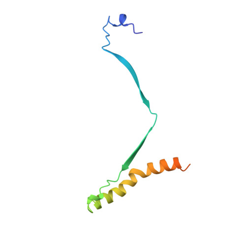
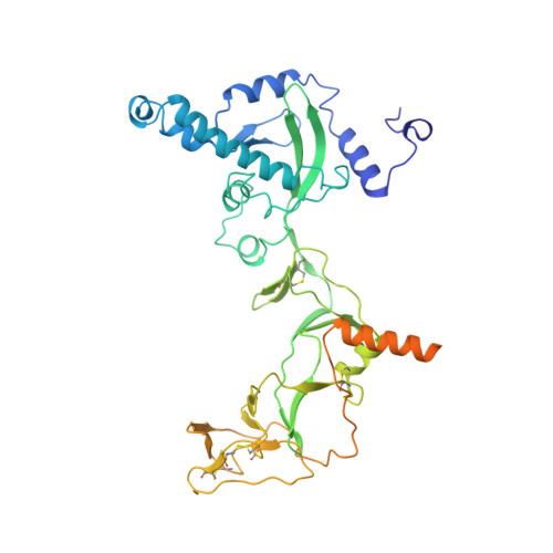
Cryo-EM structure of the prefusion state of canine distemper virus fusion protein ectodomain.

Kalbermatter, D.Shrestha, N.Gall, F.M.Wyss, M.Riedl, R.Plattet, P.Fotiadis, D.

(2020) J Struct Biol X 4: 100021-100021

  • DOI: https://doi.org/10.1016/j.yjsbx.2020.100021
  • Primary Citation of Related Structures:  
    6XYE

  • PubMed Abstract: 

    Measles virus (MeV) and canine distemper virus (CDV), two members of the Morbillivirus genus, are still causing important global diseases of humans and animals, respectively. To enter target cells, morbilliviruses rely on an envelope-anchored machinery, which is composed of two interacting glycoproteins: a tetrameric receptor binding (H) protein and a trimeric fusion (F) protein. To execute membrane fusion, the F protein initially adopts a metastable, prefusion state that refolds into a highly stable postfusion conformation as the result of a finely coordinated activation process mediated by the H protein. Here, we employed cryo-electron microscopy (cryo-EM) and single particle reconstruction to elucidate the structure of the prefusion state of the CDV F protein ectodomain (solF) at 4.3 Å resolution. Stabilization of the prefusion solF trimer was achieved by fusing the GCNt trimerization sequence at the C-terminal protein region, and expressing and purifying the recombinant protein in the presence of a morbilliviral fusion inhibitor class compound. The three-dimensional cryo-EM map of prefusion CDV solF in complex with the inhibitor clearly shows density for the ligand at the protein binding site suggesting common mechanisms of membrane fusion activation and inhibition employed by different morbillivirus members.


  • Organizational Affiliation

    Institute of Biochemistry and Molecular Medicine, University of Bern, Bern, Switzerland.


Macromolecules
Find similar proteins by:  (by identity cutoff)  |  3D Structure
Entity ID: 1
MoleculeChains Sequence LengthOrganismDetailsImage
Fusion glycoprotein F2
A, C, E
89Morbillivirus canisMutation(s): 0 
UniProt
Find proteins for A0A0R5ZPI3 (Morbillivirus canis)
Explore A0A0R5ZPI3 
Go to UniProtKB:  A0A0R5ZPI3
Entity Groups  
Sequence Clusters30% Identity50% Identity70% Identity90% Identity95% Identity100% Identity
UniProt GroupA0A0R5ZPI3
Sequence Annotations
Expand
  • Reference Sequence
Find similar proteins by:  (by identity cutoff)  |  3D Structure
Entity ID: 2
MoleculeChains Sequence LengthOrganismDetailsImage
Fusion glycoprotein F1
B, D, F
438Morbillivirus canisMutation(s): 0 
UniProt
Find proteins for Q9YKL7 (Morbillivirus canis)
Explore Q9YKL7 
Go to UniProtKB:  Q9YKL7
Entity Groups  
Sequence Clusters30% Identity50% Identity70% Identity90% Identity95% Identity100% Identity
UniProt GroupQ9YKL7
Sequence Annotations
Expand
  • Reference Sequence
Experimental Data & Validation

Experimental Data

  • Method: ELECTRON MICROSCOPY
  • Resolution: 4.30 Å
  • Aggregation State: PARTICLE 
  • Reconstruction Method: SINGLE PARTICLE 
EM Software:
TaskSoftware PackageVersion
RECONSTRUCTIONRELION3
MODEL REFINEMENTPHENIX1.16-3546

Structure Validation

View Full Validation Report


Entry History & Funding Information

Deposition Data


Funding OrganizationLocationGrant Number
Swiss National Science FoundationSwitzerlandSinergia, Ref. No. 183481

Revision History  (Full details and data files)

  • Version 1.0: 2020-07-22
    Type: Initial release