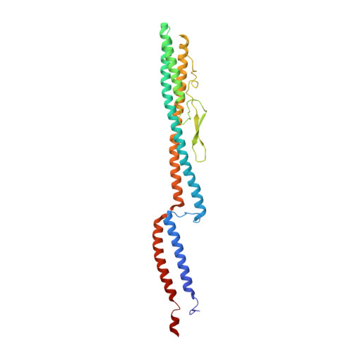
Flagellar Structures from the Bacterium Caulobacter crescentus and Implications for Phage phi CbK Predation of Multiflagellin Bacteria
Montemayor, E.J., Ploscariu, N.T., Sanchez, J.C., Parrell, D., Dillard, R.S., Shebelut, C.W., Ke, Z., Guerrero-Ferreira, R.C., Wright, E.R.(2021) J Bacteriol 203
- PubMed: 33288623
- DOI: https://doi.org/10.1128/JB.00399-20
- Primary Citation of Related Structures:
6XKY, 6XL0 - PubMed Abstract:
Caulobacter crescentus is a Gram-negative alphaproteobacterium that commonly lives in oligotrophic fresh- and saltwater environments. C. crescentus is a host to many bacteriophages, including ϕCbK and ϕCbK-like bacteriophages, which require interaction with the bacterial flagellum and pilus complexes during adsorption. It is commonly thought that the six paralogs of the flagellin gene present in C. crescentus are important for bacteriophage evasion. Here, we show that deletion of specific flagellins in C. crescentus can indeed attenuate ϕCbK adsorption efficiency, although no single deletion completely ablates ϕCbK adsorption. Thus, the bacteriophage ϕCbK likely recognizes a common motif among the six known flagellins in C. crescentus with various degrees of efficiency. Interestingly, we observe that most deletion strains still generate flagellar filaments, with the exception of a strain that contains only the most divergent flagellin, FljJ, or a strain that contains only FljN and FljO. To visualize the surface residues that are likely recognized by ϕCbK, we determined two high-resolution structures of the FljK filament, with and without an amino acid substitution that induces straightening of the filament. We observe posttranslational modifications on conserved surface threonine residues of FljK that are likely O-linked glycans. The possibility of interplay between these modifications and ϕCbK adsorption is discussed. We also determined the structure of a filament composed of a heterogeneous mixture of FljK and FljL, the final resolution of which was limited to approximately 4.6 Å. Altogether, this work builds a platform for future investigations of how phage ϕCbK infects C. crescentus at the molecular level. IMPORTANCE Bacterial flagellar filaments serve as an initial attachment point for many bacteriophages to bacteria. Some bacteria harbor numerous flagellin genes and are therefore able to generate flagellar filaments with complex compositions, which is thought to be important for evasion from bacteriophages. This study characterizes the importance of the six flagellin genes in C. crescentus for infection by bacteriophage ϕCbK. We find that filaments containing the FljK flagellin are the preferred substrate for bacteriophage ϕCbK. We also present a high-resolution structure of a flagellar filament containing only the FljK flagellin, which provides a platform for future studies on determining how bacteriophage ϕCbK attaches to flagellar filaments at the molecular level.
Organizational Affiliation:
Department of Biochemistry, University of Wisconsin, Madison, Wisconsin, USA.


