6XE9

10S myosin II (smooth muscle)

  • Classification: CONTRACTILE PROTEIN
  • Organism(s): Meleagris gallopavo
  • Mutation(s): No 

  • Deposited: 2020-06-12 Released: 2020-12-02 
  • Deposition Author(s): Tiwari, P., Craig, R., Padron, R.
  • Funding Organization(s): National Institutes of Health/National Institute of Arthritis and Musculoskeletal and Skin Diseases (NIH/NIAMS), National Institutes of Health/National Heart, Lung, and Blood Institute (NIH/NHLBI)

Experimental Data Snapshot

  • Method: ELECTRON MICROSCOPY
  • Resolution: 4.30 Å
  • Aggregation State: PARTICLE 
  • Reconstruction Method: SINGLE PARTICLE 

wwPDB Validation   3D Report Full Report


This is version 1.3 of the entry. See complete history


Literature

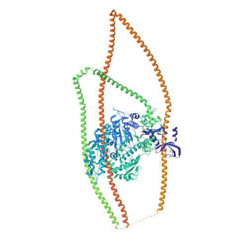
Cryo-EM structure of the inhibited (10S) form of myosin II.

Yang, S.Tiwari, P.Lee, K.H.Sato, O.Ikebe, M.Padron, R.Craig, R.

(2020) Nature 588: 521-525

  • DOI: https://doi.org/10.1038/s41586-020-3007-0
  • Primary Citation of Related Structures:  
    6XE9

  • PubMed Abstract: 

    Myosin II is the motor protein that enables muscle cells to contract and nonmuscle cells to move and change shape 1 . The molecule has two identical heads attached to an elongated tail, and can exist in two conformations: 10S and 6S, named for their sedimentation coefficients 2,3 . The 6S conformation has an extended tail and assembles into polymeric filaments, which pull on actin filaments to generate force and motion. In 10S myosin, the tail is folded into three segments and the heads bend back and interact with each other and the tail 3-7 , creating a compact conformation in which ATPase activity, actin activation and filament assembly are all highly inhibited 7,8 . This switched-off structure appears to function as a key energy-conserving storage molecule in muscle and nonmuscle cells 9-12 , which can be activated to form functional filaments as needed 13 -but the mechanism of its inhibition is not understood. Here we have solved the structure of smooth muscle 10S myosin by cryo-electron microscopy with sufficient resolution to enable improved understanding of the function of the head and tail regions of the molecule and of the key intramolecular contacts that cause inhibition. Our results suggest an atomic model for the off state of myosin II, for its activation and unfolding by phosphorylation, and for understanding the clustering of disease-causing mutations near sites of intramolecular interaction.


  • Organizational Affiliation

    Division of Cell Biology and Imaging, Department of Radiology, University of Massachusetts Medical School, Worcester, MA, USA.


Macromolecules
Find similar proteins by:  (by identity cutoff)  |  3D Structure
Entity ID: 1
MoleculeChains Sequence LengthOrganismDetailsImage
Myosin II heavy chain (smooth muscle)A,
D [auth M]
1,979Meleagris gallopavoMutation(s): 0 
EC: 5.6.1.8
UniProt
Find proteins for G1N5L2 (Meleagris gallopavo)
Explore G1N5L2 
Go to UniProtKB:  G1N5L2
Entity Groups  
Sequence Clusters30% Identity50% Identity70% Identity90% Identity95% Identity100% Identity
UniProt GroupG1N5L2
Sequence Annotations
Expand
  • Reference Sequence
Find similar proteins by:  (by identity cutoff)  |  3D Structure
Entity ID: 2
MoleculeChains Sequence LengthOrganismDetailsImage
Myosin light chain smooth muscle isoformB,
E [auth N]
151Meleagris gallopavoMutation(s): 0 
UniProt
Find proteins for Q6W5H0 (Meleagris gallopavo)
Explore Q6W5H0 
Go to UniProtKB:  Q6W5H0
Entity Groups  
Sequence Clusters30% Identity50% Identity70% Identity90% Identity95% Identity100% Identity
UniProt GroupQ6W5H0
Sequence Annotations
Expand
  • Reference Sequence
Find similar proteins by:  (by identity cutoff)  |  3D Structure
Entity ID: 3
MoleculeChains Sequence LengthOrganismDetailsImage
Myosin light chain 9C,
F [auth O]
172Meleagris gallopavoMutation(s): 0 
UniProt
Find proteins for G3URE9 (Meleagris gallopavo)
Explore G3URE9 
Go to UniProtKB:  G3URE9
Entity Groups  
Sequence Clusters30% Identity50% Identity70% Identity90% Identity95% Identity100% Identity
UniProt GroupG3URE9
Sequence Annotations
Expand
  • Reference Sequence
Experimental Data & Validation

Experimental Data

  • Method: ELECTRON MICROSCOPY
  • Resolution: 4.30 Å
  • Aggregation State: PARTICLE 
  • Reconstruction Method: SINGLE PARTICLE 
EM Software:
TaskSoftware PackageVersion
RECONSTRUCTIONRELION3.0
MODEL REFINEMENTCoot0.8.9.2 EL
MODEL REFINEMENTPHENIX1.17.1 - 3660

Structure Validation

View Full Validation Report


Entry History & Funding Information

Deposition Data


Funding OrganizationLocationGrant Number
National Institutes of Health/National Institute of Arthritis and Musculoskeletal and Skin Diseases (NIH/NIAMS)United StatesR01AR072036
National Institutes of Health/National Institute of Arthritis and Musculoskeletal and Skin Diseases (NIH/NIAMS)United StatesR01AR067279
National Institutes of Health/National Heart, Lung, and Blood Institute (NIH/NHLBI)United StatesR01HL139883

Revision History  (Full details and data files)

  • Version 1.0: 2020-12-02
    Type: Initial release
  • Version 1.1: 2020-12-16
    Changes: Database references
  • Version 1.2: 2020-12-30
    Changes: Database references
  • Version 1.3: 2024-03-06
    Changes: Data collection, Database references, Refinement description