6XB5

Structure of Trichoplusia ni poxin in post-reactive state with Gp[2'-5']Ap[3']


Experimental Data Snapshot

  • Method: X-RAY DIFFRACTION
  • Resolution: 2.90 Å
  • R-Value Free: 0.215 
  • R-Value Work: 0.184 
  • R-Value Observed: 0.187 

wwPDB Validation   3D Report Full Report


Ligand Structure Quality Assessment 


This is version 1.1 of the entry. See complete history


Literature

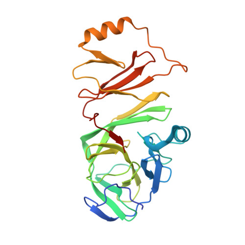
Structures of diverse poxin cGAMP nucleases reveal a widespread role for cGAS-STING evasion in host-pathogen conflict.

Eaglesham, J.B.McCarty, K.L.Kranzusch, P.J.

(2020) Elife 9

  • DOI: https://doi.org/10.7554/eLife.59753
  • Primary Citation of Related Structures:  
    6XB3, 6XB4, 6XB5, 6XB6

  • PubMed Abstract: 

    DNA viruses in the family Poxviridae encode poxin enzymes that degrade the immune second messenger 2'3'-cGAMP to inhibit cGAS-STING immunity in mammalian cells. The closest homologs of poxin exist in the genomes of insect viruses suggesting a key mechanism of cGAS-STING evasion may have evolved outside of mammalian biology. Here we use a biochemical and structural approach to discover a broad family of 369 poxins encoded in diverse viral and animal genomes and define a prominent role for 2'3'-cGAMP cleavage in metazoan host-pathogen conflict. Structures of insect poxins reveal unexpected homology to flavivirus proteases and enable identification of functional self-cleaving poxins in RNA-virus polyproteins. Our data suggest widespread 2'3'-cGAMP signaling in insect antiviral immunity and explain how a family of cGAS-STING evasion enzymes evolved from viral proteases through gain of secondary nuclease activity. Poxin acquisition by poxviruses demonstrates the importance of environmental connections in shaping evolution of mammalian pathogens.


  • Organizational Affiliation

    Department of Microbiology, Harvard Medical School, Boston, United States.


Macromolecules
Find similar proteins by:  (by identity cutoff)  |  3D Structure
Entity ID: 1
MoleculeChains Sequence LengthOrganismDetailsImage
Poxin
A, B
244Trichoplusia niMutation(s): 0 
UniProt
Find proteins for A0A7E5VPD6 (Trichoplusia ni)
Explore A0A7E5VPD6 
Go to UniProtKB:  A0A7E5VPD6
Entity Groups  
Sequence Clusters30% Identity50% Identity70% Identity90% Identity95% Identity100% Identity
UniProt GroupA0A7E5VPD6
Sequence Annotations
Expand
  • Reference Sequence
Experimental Data & Validation

Experimental Data

  • Method: X-RAY DIFFRACTION
  • Resolution: 2.90 Å
  • R-Value Free: 0.215 
  • R-Value Work: 0.184 
  • R-Value Observed: 0.187 
  • Space Group: I 41
Unit Cell:
Length ( Å )Angle ( ˚ )
a = 124.422α = 90
b = 124.422β = 90
c = 132.178γ = 90
Software Package:
Software NamePurpose
PHENIXrefinement
XDSdata reduction
Aimlessdata scaling
PHASERphasing

Structure Validation

View Full Validation Report


Ligand Structure Quality Assessment 


Entry History 

Deposition Data

Revision History  (Full details and data files)

  • Version 1.0: 2020-11-25
    Type: Initial release
  • Version 1.1: 2023-10-18
    Changes: Data collection, Database references, Refinement description