6WXV

CryoEM structure of mouse DUOX1-DUOXA1 complex in the presence of NADPH


Experimental Data Snapshot

  • Method: ELECTRON MICROSCOPY
  • Resolution: 3.30 Å
  • Aggregation State: PARTICLE 
  • Reconstruction Method: SINGLE PARTICLE 

wwPDB Validation   3D Report Full Report


This is version 1.1 of the entry. See complete history


Literature

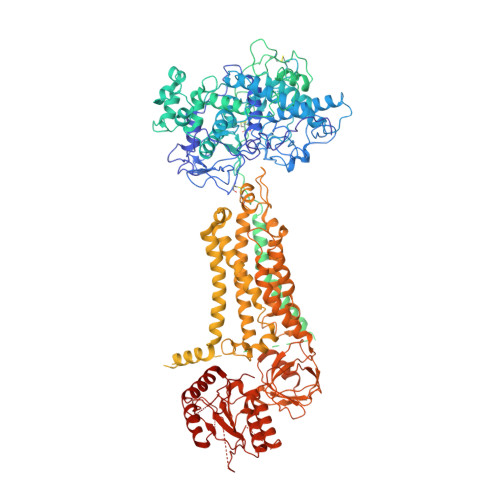
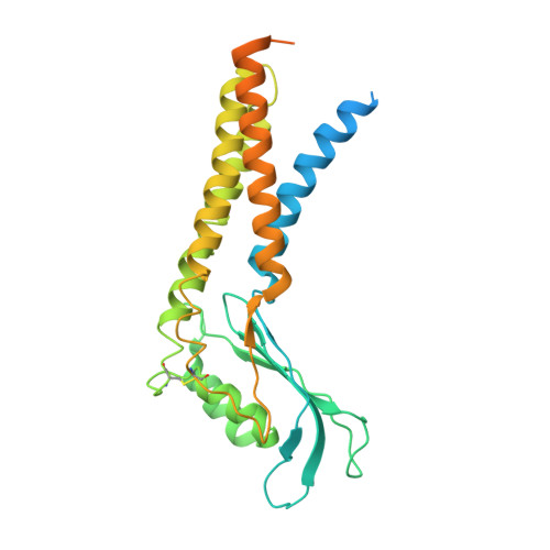
Structures of mouse DUOX1-DUOXA1 provide mechanistic insights into enzyme activation and regulation.

Sun, J.

(2020) Nat Struct Mol Biol 27: 1086-1093

  • DOI: https://doi.org/10.1038/s41594-020-0501-x
  • Primary Citation of Related Structures:  
    6WXR, 6WXU, 6WXV

  • PubMed Abstract: 

    DUOX1, an NADPH oxidase family member, catalyzes the production of hydrogen peroxide. DUOX1 is expressed in various tissues, including the thyroid and respiratory tract, and plays a crucial role in processes such as thyroid hormone biosynthesis and innate host defense. DUOX1 co-assembles with its maturation factor DUOXA1 to form an active enzyme complex. However, the molecular mechanisms for activation and regulation of DUOX1 remain mostly unclear. Here, I present cryo-EM structures of the mammalian DUOX1-DUOXA1 complex, in the absence and presence of substrate NADPH, as well as DUOX1-DUOXA1 in an unexpected dimer-of-dimers configuration. These structures reveal atomic details of the DUOX1-DUOXA1 interaction, a lipid-mediated NADPH-binding pocket and the electron transfer path. Furthermore, biochemical and structural analyses indicate that the dimer-of-dimers configuration represents an inactive state of DUOX1-DUOXA1, suggesting an oligomerization-dependent regulatory mechanism. Together, my work provides structural bases for DUOX1-DUOXA1 activation and regulation.


  • Organizational Affiliation

    Department of Structural Biology, St Jude Children's Research Hospital, Memphis, TN, USA. ji.sun@stjude.org.


Macromolecules
Find similar proteins by:  (by identity cutoff)  |  3D Structure
Entity ID: 1
MoleculeChains Sequence LengthOrganismDetailsImage
Dual oxidase 11,536Mus musculusMutation(s): 0 
Gene Names: Duox1
Membrane Entity: Yes 
UniProt
Find proteins for A2AQ92 (Mus musculus)
Explore A2AQ92 
Go to UniProtKB:  A2AQ92
Entity Groups  
Sequence Clusters30% Identity50% Identity70% Identity90% Identity95% Identity100% Identity
UniProt GroupA2AQ92
Sequence Annotations
Expand
  • Reference Sequence
Find similar proteins by:  (by identity cutoff)  |  3D Structure
Entity ID: 2
MoleculeChains Sequence LengthOrganismDetailsImage
Dual oxidase maturation factor 1341Mus musculusMutation(s): 0 
Gene Names: Duoxa1
Membrane Entity: Yes 
UniProt
Find proteins for Q8VE49 (Mus musculus)
Explore Q8VE49 
Go to UniProtKB:  Q8VE49
Entity Groups  
Sequence Clusters30% Identity50% Identity70% Identity90% Identity95% Identity100% Identity
UniProt GroupQ8VE49
Sequence Annotations
Expand
  • Reference Sequence
Oligosaccharides

Help

Entity ID: 3
MoleculeChains Length2D Diagram Glycosylation3D Interactions
alpha-D-mannopyranose-(1-3)-alpha-D-mannopyranose-(1-6)-beta-D-mannopyranose-(1-4)-2-acetamido-2-deoxy-beta-D-glucopyranose-(1-4)-2-acetamido-2-deoxy-beta-D-glucopyranose
C
5N-Glycosylation
Glycosylation Resources
GlyTouCan:  G10756ZZ
GlyCosmos:  G10756ZZ
GlyGen:  G10756ZZ
Small Molecules
Ligands 6 Unique
IDChains Name / Formula / InChI Key2D Diagram3D Interactions
FAD
Query on FAD

Download Ideal Coordinates CCD File 
D [auth A]FLAVIN-ADENINE DINUCLEOTIDE
C27 H33 N9 O15 P2
VWWQXMAJTJZDQX-UYBVJOGSSA-N
NDP (Subject of Investigation/LOI)
Query on NDP

Download Ideal Coordinates CCD File 
J [auth A]NADPH DIHYDRO-NICOTINAMIDE-ADENINE-DINUCLEOTIDE PHOSPHATE
C21 H30 N7 O17 P3
ACFIXJIJDZMPPO-NNYOXOHSSA-N
PLC (Subject of Investigation/LOI)
Query on PLC

Download Ideal Coordinates CCD File 
K [auth A]DIUNDECYL PHOSPHATIDYL CHOLINE
C32 H65 N O8 P
IJFVSSZAOYLHEE-SSEXGKCCSA-O
HEB
Query on HEB

Download Ideal Coordinates CCD File 
H [auth A],
I [auth A]
HEME B/C
C34 H34 Fe N4 O4
NEGHHAJBRZGUAY-RGGAHWMASA-L
NAG
Query on NAG

Download Ideal Coordinates CCD File 
E [auth A],
F [auth A],
G [auth A],
M [auth B]
2-acetamido-2-deoxy-beta-D-glucopyranose
C8 H15 N O6
OVRNDRQMDRJTHS-FMDGEEDCSA-N
CA
Query on CA

Download Ideal Coordinates CCD File 
L [auth A]CALCIUM ION
Ca
BHPQYMZQTOCNFJ-UHFFFAOYSA-N
Experimental Data & Validation

Experimental Data

  • Method: ELECTRON MICROSCOPY
  • Resolution: 3.30 Å
  • Aggregation State: PARTICLE 
  • Reconstruction Method: SINGLE PARTICLE 
EM Software:
TaskSoftware PackageVersion
RECONSTRUCTIONcryoSPARC2.13

Structure Validation

View Full Validation Report


Entry History & Funding Information

Deposition Data

  • Released Date: 2020-09-02 
  • Deposition Author(s): Sun, J.

Funding OrganizationLocationGrant Number
National Institutes of Health/National Heart, Lung, and Blood Institute (NIH/NHLBI)United StatesHL143037

Revision History  (Full details and data files)

  • Version 1.0: 2020-09-02
    Type: Initial release
  • Version 1.1: 2021-03-17
    Changes: Database references