6WT4

Structure of a bacterial STING receptor from Flavobacteriaceae sp. in complex with 3',3'-cGAMP


Experimental Data Snapshot

  • Method: X-RAY DIFFRACTION
  • Resolution: 1.78 Å
  • R-Value Free: 0.230 
  • R-Value Work: 0.199 
  • R-Value Observed: 0.200 

wwPDB Validation   3D Report Full Report


Ligand Structure Quality Assessment 


This is version 1.2 of the entry. See complete history


Literature

STING cyclic dinucleotide sensing originated in bacteria.

Morehouse, B.R.Govande, A.A.Millman, A.Keszei, A.F.A.Lowey, B.Ofir, G.Shao, S.Sorek, R.Kranzusch, P.J.

(2020) Nature 586: 429-433

  • DOI: https://doi.org/10.1038/s41586-020-2719-5
  • Primary Citation of Related Structures:  
    6WT4, 6WT5, 6WT6, 6WT7, 6WT8, 6WT9

  • PubMed Abstract: 

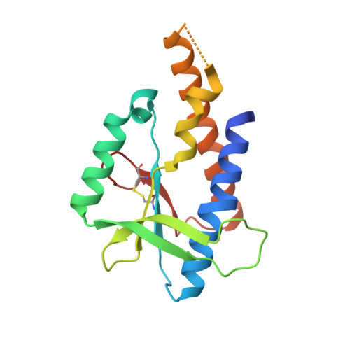
    Stimulator of interferon genes (STING) is a receptor in human cells that senses foreign cyclic dinucleotides that are released during bacterial infection and in endogenous cyclic GMP-AMP signalling during viral infection and anti-tumour immunity 1-5 . STING shares no structural homology with other known signalling proteins 6-9 , which has limited attempts at functional analysis and prevented explanation of the origin of cyclic dinucleotide signalling in mammalian innate immunity. Here we reveal functional STING homologues encoded within prokaryotic defence islands, as well as a conserved mechanism of signal activation. Crystal structures of bacterial STING define a minimal homodimeric scaffold that selectively responds to cyclic di-GMP synthesized by a neighbouring cGAS/DncV-like nucleotidyltransferase (CD-NTase) enzyme. Bacterial STING domains couple the recognition of cyclic dinucleotides with the formation of protein filaments to drive oligomerization of TIR effector domains and rapid NAD + cleavage. We reconstruct the evolutionary events that followed the acquisition of STING into metazoan innate immunity, and determine the structure of a full-length TIR-STING fusion from the Pacific oyster Crassostrea gigas. Comparative structural analysis demonstrates how metazoan-specific additions to the core STING scaffold enabled a switch from direct effector function to regulation of antiviral transcription. Together, our results explain the mechanism of STING-dependent signalling and reveal the conservation of a functional cGAS-STING pathway in prokaryotic defence against bacteriophages.


  • Organizational Affiliation

    Department of Microbiology, Harvard Medical School, Boston, MA, USA.


Macromolecules
Find similar proteins by:  (by identity cutoff)  |  3D Structure
Entity ID: 1
MoleculeChains Sequence LengthOrganismDetailsImage
Bacterial STING
A, B
162FlavobacteriaceaeMutation(s): 0 
UniProt
Find proteins for P0DUD7 (Flavobacteriaceae sp. genome_bin_11)
Explore P0DUD7 
Go to UniProtKB:  P0DUD7
Entity Groups  
Sequence Clusters30% Identity50% Identity70% Identity90% Identity95% Identity100% Identity
UniProt GroupP0DUD7
Sequence Annotations
Expand
  • Reference Sequence
Small Molecules
Ligands 2 Unique
IDChains Name / Formula / InChI Key2D Diagram3D Interactions
4BW (Subject of Investigation/LOI)
Query on 4BW

Download Ideal Coordinates CCD File 
C [auth A]2-amino-9-[(2R,3R,3aS,5R,7aR,9R,10R,10aS,12R,14aR)-9-(6-amino-9H-purin-9-yl)-3,5,10,12-tetrahydroxy-5,12-dioxidooctahydro-2H,7H-difuro[3,2-d:3',2'-j][1,3,7,9,2,8]tetraoxadiphosphacyclododecin-2-yl]-1,9-dihydro-6H-purin-6-one
C20 H24 N10 O13 P2
RFCBNSCSPXMEBK-INFSMZHSSA-N
SO4
Query on SO4

Download Ideal Coordinates CCD File 
D [auth B],
E [auth B]
SULFATE ION
O4 S
QAOWNCQODCNURD-UHFFFAOYSA-L
Experimental Data & Validation

Experimental Data

  • Method: X-RAY DIFFRACTION
  • Resolution: 1.78 Å
  • R-Value Free: 0.230 
  • R-Value Work: 0.199 
  • R-Value Observed: 0.200 
  • Space Group: I 4
Unit Cell:
Length ( Å )Angle ( ˚ )
a = 121.848α = 90
b = 121.848β = 90
c = 48.16γ = 90
Software Package:
Software NamePurpose
PHENIXrefinement
XDSdata reduction
Aimlessdata scaling
AutoSolphasing

Structure Validation

View Full Validation Report


Ligand Structure Quality Assessment 


Entry History 

Deposition Data

Revision History  (Full details and data files)

  • Version 1.0: 2020-09-09
    Type: Initial release
  • Version 1.1: 2020-09-16
    Changes: Database references, Source and taxonomy, Structure summary
  • Version 1.2: 2020-10-28
    Changes: Database references