CryoEM structure of the low-complexity domain of hnRNPA2 and its conversion to pathogenic amyloid.
Lu, J., Cao, Q., Hughes, M.P., Sawaya, M.R., Boyer, D.R., Cascio, D., Eisenberg, D.S.(2020) Nat Commun 11: 4090-4090
- PubMed: 32796831
- DOI: https://doi.org/10.1038/s41467-020-17905-y
- Primary Citation of Related Structures:
6WPQ, 6WQK - PubMed Abstract:
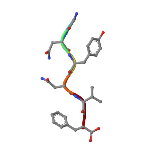
hnRNPA2 is a human ribonucleoprotein (RNP) involved in RNA metabolism. It forms fibrils both under cellular stress and in mutated form in neurodegenerative conditions. Previous work established that the C-terminal low-complexity domain (LCD) of hnRNPA2 fibrillizes under stress, and missense mutations in this domain are found in the disease multisystem proteinopathy (MSP). However, little is known at the atomic level about the hnRNPA2 LCD structure that is involved in those processes and how disease mutations cause structural change. Here we present the cryo-electron microscopy (cryoEM) structure of the hnRNPA2 LCD fibril core and demonstrate its capability to form a reversible hydrogel in vitro containing amyloid-like fibrils. Whereas these fibrils, like pathogenic amyloid, are formed from protein chains stacked into β-sheets by backbone hydrogen bonds, they display distinct structural differences: the chains are kinked, enabling non-covalent cross-linking of fibrils and disfavoring formation of pathogenic steric zippers. Both reversibility and energetic calculations suggest these fibrils are less stable than pathogenic amyloid. Moreover, the crystal structure of the disease-mutation-containing segment (D290V) of hnRNPA2 suggests that the replacement fundamentally alters the fibril structure to a more stable energetic state. These findings illuminate how molecular interactions promote protein fibril networks and how mutation can transform fibril structure from functional to a pathogenic form.
Organizational Affiliation:
Departments of Chemistry and Biochemistry and Biological Chemistry, University of California, Los Angeles, Los Angeles, CA, USA.