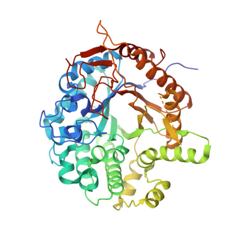
X-ray Structure, Bioinformatics Analysis, and Substrate Specificity of a 6-Phospho-beta-glucosidase Glycoside Hydrolase 1 Enzyme from Bacillus licheniformis .
Veldman, W., Liberato, M.V., Almeida, V.M., Souza, V.P., Frutuoso, M.A., Marana, S.R., Moses, V., Tastan Bishop, O., Polikarpov, I.(2020) J Chem Inf Model 60: 6392-6407
- PubMed: 33166469
- DOI: https://doi.org/10.1021/acs.jcim.0c00759
- Primary Citation of Related Structures:
6WGD - PubMed Abstract:
In bacteria, mono- and disaccharides are phosphorylated during the uptake processes through the vastly spread transport system phosphoenolpyruvate-dependent phosphotransferase. As an initial step in the phosphorylated disaccharide metabolism pathway, 6-phospho-β-glucosidases and 6-phospho-β-galactosidases play a crucial role by releasing phosphorylated and nonphosphorylated monosaccharides. However, structural determinants for the specificity of these enzymes still need to be clarified. Here, an X-ray structure of a glycoside hydrolase family 1 enzyme from Bacillus licheniformis , hereafter known as Bl BglH, was determined at 2.2 Å resolution, and its substrate specificity was investigated. The sequence of Bl BglH was compared to the sequences of 58 other GH1 enzymes using sequence alignments, sequence identity calculations, phylogenetic analysis, and motif discovery. Through these various analyses, Bl BglH was found to have sequence features characteristic of the 6-phospho-β-glucosidase activity enzymes. Motif and structural observations highlighted the importance of loop L8 in 6-phospho-β-glucosidase activity enzymes. To further affirm enzyme specificity, molecular docking and molecular dynamics simulations were performed using the crystallographic structure of Bl BglH. Docking was carried out with a 6-phospho-β-glucosidase enzyme activity positive and negative control ligand, followed by 400 ns of MD simulations. The positive and negative control ligands were PNP6Pglc and PNP6Pgal, respectively. PNP6Pglc maintained favorable interactions within the active site until the end of the MD simulation, while PNP6Pgal exhibited instability. The favorable binding of substrate stabilized the loops that surround the active site. Binding free energy calculations showed that the PNP6Pglc complex had a substantially lower binding energy compared to the PNP6Pgal complex. Altogether, the findings of this study suggest that Bl BglH possesses 6-phospho-β-glucosidase enzymatic activity and revealed sequence and structural differences between bacterial GH1 enzymes of various activities.
Organizational Affiliation:
Research Unit in Bioinformatics (RUBi), Department of Biochemistry and Microbiology, Rhodes University, Grahamstown 6140, South Africa.


