Structural insight into the novel iron-coordination and domain interactions of transferrin-1 from a model insect, Manduca sexta.
Weber, J.J., Kashipathy, M.M., Battaile, K.P., Go, E., Desaire, H., Kanost, M.R., Lovell, S., Gorman, M.J.(2021) Protein Sci 30: 408-422
- PubMed: 33197096
- DOI: https://doi.org/10.1002/pro.3999
- Primary Citation of Related Structures:
6WB6 - PubMed Abstract:
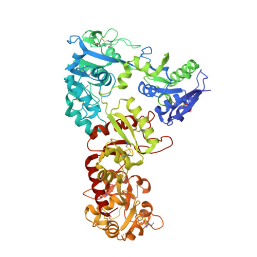
Transferrins function in iron sequestration and iron transport by binding iron tightly and reversibly. Vertebrate transferrins coordinate iron through interactions with two tyrosines, an aspartate, a histidine, and a carbonate anion, and conformational changes that occur upon iron binding and release have been described. Much less is known about the structure and functions of insect transferrin-1 (Tsf1), which is present in hemolymph and influences iron homeostasis mostly by unknown mechanisms. Amino acid sequence and biochemical analyses have suggested that iron coordination by Tsf1 differs from that of the vertebrate transferrins. Here we report the first crystal structure (2.05 Å resolution) of an insect transferrin. Manduca sexta (MsTsf1) in the holo form exhibits a bilobal fold similar to that of vertebrate transferrins, but its carboxyl-lobe adopts a novel orientation and contacts with the amino-lobe. The structure revealed coordination of a single Fe 3+ ion in the amino-lobe through Tyr90, Tyr204, and two carbonate anions. One carbonate anion is buried near the ferric ion and is coordinated by four residues, whereas the other carbonate anion is solvent exposed and coordinated by Asn121. Notably, these residues are highly conserved in Tsf1 orthologs. Docking analysis suggested that the solvent exposed carbonate position is capable of binding alternative anions. These findings provide a structural basis for understanding Tsf1 function in iron sequestration and transport in insects as well as insight into the similarities and differences in iron homeostasis between insects and humans.
Organizational Affiliation:
Department of Biochemistry and Molecular Biophysics, Kansas State University, Manhattan, Kansas, USA.