6W5Y

Inferred receptor binding domain of human endogenous retrovirus envelope EnvP(b)1

  • Classification: VIRAL PROTEIN
  • Organism(s): Gammaretrovirus
  • Expression System: Homo sapiens
  • Mutation(s): No 

  • Deposited: 2020-03-14 Released: 2020-11-11 
  • Deposition Author(s): McCarthy, K.R.
  • Funding Organization(s): National Institutes of Health/National Institute Of Allergy and Infectious Diseases (NIH/NIAID), Howard Hughes Medical Institute (HHMI)

Experimental Data Snapshot

  • Method: X-RAY DIFFRACTION
  • Resolution: 2.50 Å
  • R-Value Free: 0.254 
  • R-Value Work: 0.222 
  • R-Value Observed: 0.224 

wwPDB Validation   3D Report Full Report


This is version 1.1 of the entry. See complete history


Literature

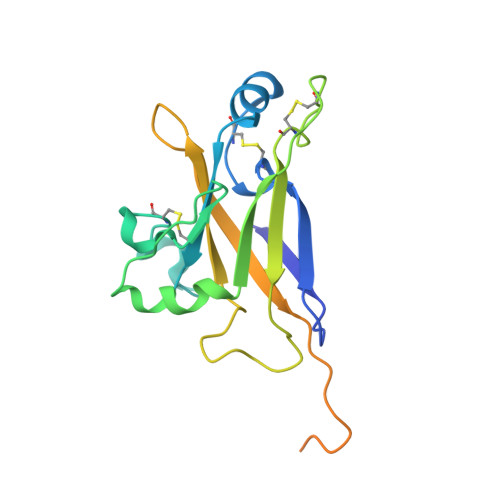
Structure of the Receptor Binding Domain of EnvP(b)1, an Endogenous Retroviral Envelope Protein Expressed in Human Tissues.

McCarthy, K.R.Timpona, J.L.Jenni, S.Bloyet, L.M.Brusic, V.Johnson, W.E.Whelan, S.P.J.Robinson-McCarthy, L.R.

(2020) mBio 11

  • DOI: https://doi.org/10.1128/mBio.02772-20
  • Primary Citation of Related Structures:  
    6W5Y

  • PubMed Abstract: 

    EnvP(b)1 is an endogenous retroviral envelope gene found in human and other primate genomes. We report EnvP(b)1 sequences in primate genomes consistent with an integration event between 40 and 71 million years ago. Using a highly specific polyclonal antiserum raised against the putative receptor binding domain (RBD) of human EnvP(b)1, we detected expression in human placenta, ovaries, and thymus. We found that EnvP(b)1 is proteolytically processed, and using cell-cell fusion assays in multiple primate cell lines, we demonstrated that extant EnvP(b)1 proteins from a variety of primate genomes are fusogenic. This work supports the idea that EnvP(b)1 is under purifying selection and its fusogenic activity has been maintained for over 40 million years. We determined the structure of the RBD of human EnvP(b)1, which defines structural similarities with extant leukemia viruses, despite little sequence conservation. This structure highlights a common scaffold from which novel receptor binding specificities likely evolved. The evolutionary plasticity of this domain may underlie the diversity of related Envs in circulating viruses. IMPORTANCE Organisms can access genetic and functional novelty by capturing viral elements within their genomes, where they can evolve to drive new cellular or organismal processes. We demonstrate that a retroviral envelope gene, EnvP(b)1, has been maintained and its fusion activity preserved for 40 to 71 million years. It is expressed as a protein in multiple healthy human tissues. We determined the structure of its inferred receptor binding domain and compared it with the same domain in modern viruses. We found a common conserved architecture that underlies the varied receptor binding activity of divergent Env genes. The modularity and versatility of this domain may underpin the evolutionary success of this clade of fusogens.


  • Organizational Affiliation

    Department of Biological Chemistry and Molecular Pharmacology, Harvard Medical School, Boston, Massachusetts, USA.


Macromolecules
Find similar proteins by:  (by identity cutoff)  |  3D Structure
Entity ID: 1
MoleculeChains Sequence LengthOrganismDetailsImage
EnvP(b)1 inferred receptor binding domain
A, B, C
201GammaretrovirusMutation(s): 0 
Gene Names: env
Entity Groups  
Sequence Clusters30% Identity50% Identity70% Identity90% Identity95% Identity100% Identity
Sequence Annotations
Expand
  • Reference Sequence
Oligosaccharides

Help

Entity ID: 2
MoleculeChains Length2D Diagram Glycosylation3D Interactions
beta-D-mannopyranose-(1-4)-2-acetamido-2-deoxy-beta-D-glucopyranose-(1-4)-2-acetamido-2-deoxy-beta-D-glucopyranose
D
3N-Glycosylation
Glycosylation Resources
GlyTouCan:  G15407YE
GlyCosmos:  G15407YE
GlyGen:  G15407YE
Entity ID: 3
MoleculeChains Length2D Diagram Glycosylation3D Interactions
2-acetamido-2-deoxy-beta-D-glucopyranose-(1-4)-[alpha-L-fucopyranose-(1-6)]2-acetamido-2-deoxy-beta-D-glucopyranose
E
3N-Glycosylation
Glycosylation Resources
GlyTouCan:  G21290RB
GlyCosmos:  G21290RB
GlyGen:  G21290RB
Entity ID: 4
MoleculeChains Length2D Diagram Glycosylation3D Interactions
2-acetamido-2-deoxy-beta-D-glucopyranose-(1-4)-2-acetamido-2-deoxy-beta-D-glucopyranose
F
2N-Glycosylation
Glycosylation Resources
GlyTouCan:  G42666HT
GlyCosmos:  G42666HT
GlyGen:  G42666HT
Experimental Data & Validation

Experimental Data

  • Method: X-RAY DIFFRACTION
  • Resolution: 2.50 Å
  • R-Value Free: 0.254 
  • R-Value Work: 0.222 
  • R-Value Observed: 0.224 
  • Space Group: C 1 2 1
Unit Cell:
Length ( Å )Angle ( ˚ )
a = 243.31α = 90
b = 37.35β = 102.28
c = 66.99γ = 90
Software Package:
Software NamePurpose
XDSdata reduction
PHENIXrefinement
PDB_EXTRACTdata extraction
XSCALEdata scaling
PHASERphasing

Structure Validation

View Full Validation Report


Entry History & Funding Information

Deposition Data


Funding OrganizationLocationGrant Number
National Institutes of Health/National Institute Of Allergy and Infectious Diseases (NIH/NIAID)United StatesAI081842
National Institutes of Health/National Institute Of Allergy and Infectious Diseases (NIH/NIAID)United StatesAI109740
Howard Hughes Medical Institute (HHMI)United States--
National Institutes of Health/National Institute Of Allergy and Infectious Diseases (NIH/NIAID)United StatesAI059371

Revision History  (Full details and data files)

  • Version 1.0: 2020-11-11
    Type: Initial release
  • Version 1.1: 2021-05-26
    Changes: Database references