6VKM

Crystal Structure of Stabilized GP from Makona Variant of Ebola Virus


Experimental Data Snapshot

  • Method: X-RAY DIFFRACTION
  • Resolution: 3.50 Å
  • R-Value Free: 0.303 
  • R-Value Work: 0.284 
  • R-Value Observed: 0.285 

wwPDB Validation   3D Report Full Report

Currently 6VKM does not have a validation slider image.


This is version 2.1 of the entry. See complete history


Literature

Structure-Based Design of Prefusion-Stabilized Filovirus Glycoprotein Trimers.

Rutten, L.Gilman, M.S.A.Blokland, S.Juraszek, J.McLellan, J.S.Langedijk, J.P.M.

(2020) Cell Rep 30: 4540-4550.e3

  • DOI: https://doi.org/10.1016/j.celrep.2020.03.025
  • Primary Citation of Related Structures:  
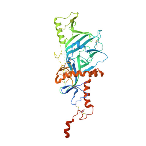
    6VKM

  • PubMed Abstract: 

    Ebola virus causes severe hemorrhagic fever, often leading to death in humans. The trimeric fusion glycoprotein (GP) is the sole target for neutralizing antibodies and is the major focus of vaccine development. Soluble GP ectodomains are unstable and mostly monomeric when not fused to a heterologous trimerization domain. Here, we report structure-based designs of Ebola and Marburg GP trimers based on a stabilizing mutation in the hinge loop in refolding region 1 and substitution of a partially buried charge at the interface of the GP1 and GP2 subunits. The combined substitutions (T577P and K588F) substantially increased trimer expression for Ebola GP proteins. We determined the crystal structure of stabilized GP from the Makona Zaire ebolavirus strain without a trimerization domain or complexed ligand. The structure reveals that the stabilized GP adopts the same trimeric prefusion conformation, provides insight into triggering of GP conformational changes, and should inform future filovirus vaccine development.


  • Organizational Affiliation

    Janssen Vaccines & Prevention, Archimedesweg 4-6, Leiden 2333 CN, the Netherlands.


Macromolecules
Find similar proteins by:  (by identity cutoff)  |  3D Structure
Entity ID: 1
MoleculeChains Sequence LengthOrganismDetailsImage
Virion spike glycoprotein494Zaire ebolavirusMutation(s): 3 
Gene Names: GP
UniProt
Find proteins for Q05320 (Zaire ebolavirus (strain Mayinga-76))
Explore Q05320 
Go to UniProtKB:  Q05320
Entity Groups  
Sequence Clusters30% Identity50% Identity70% Identity90% Identity95% Identity100% Identity
UniProt GroupQ05320
Sequence Annotations
Expand
  • Reference Sequence
Oligosaccharides

Help

Entity ID: 2
MoleculeChains Length2D Diagram Glycosylation3D Interactions
2-acetamido-2-deoxy-beta-D-glucopyranose-(1-4)-2-acetamido-2-deoxy-beta-D-glucopyranose
B
2N-Glycosylation
Glycosylation Resources
GlyTouCan:  G42666HT
GlyCosmos:  G42666HT
GlyGen:  G42666HT
Small Molecules
Ligands 1 Unique
IDChains Name / Formula / InChI Key2D Diagram3D Interactions
NAG
Query on NAG

Download Ideal Coordinates CCD File 
C [auth A]2-acetamido-2-deoxy-beta-D-glucopyranose
C8 H15 N O6
OVRNDRQMDRJTHS-FMDGEEDCSA-N
Experimental Data & Validation

Experimental Data

  • Method: X-RAY DIFFRACTION
  • Resolution: 3.50 Å
  • R-Value Free: 0.303 
  • R-Value Work: 0.284 
  • R-Value Observed: 0.285 
  • Space Group: H 3 2
Unit Cell:
Length ( Å )Angle ( ˚ )
a = 113.11α = 90
b = 113.11β = 90
c = 393.18γ = 120
Software Package:
Software NamePurpose
Aimlessdata scaling
PHENIXrefinement
PDB_EXTRACTdata extraction
iMOSFLMdata reduction
PHASERphasing

Structure Validation

View Full Validation Report

Currently 6VKM does not have a validation slider image.


Entry History 

Deposition Data

Revision History  (Full details and data files)

  • Version 1.0: 2020-04-15
    Type: Initial release
  • Version 2.0: 2020-07-29
    Type: Remediation
    Reason: Carbohydrate remediation
    Changes: Atomic model, Data collection, Derived calculations, Structure summary
  • Version 2.1: 2023-10-11
    Changes: Data collection, Database references, Refinement description, Structure summary