6VJZ

CryoEM structure of Hrd1-Usa1/Der1/Hrd3 complex of the expected topology


Experimental Data Snapshot

  • Method: ELECTRON MICROSCOPY
  • Resolution: 4.30 Å
  • Aggregation State: PARTICLE 
  • Reconstruction Method: SINGLE PARTICLE 

wwPDB Validation   3D Report Full Report


This is version 1.2 of the entry. See complete history


Literature

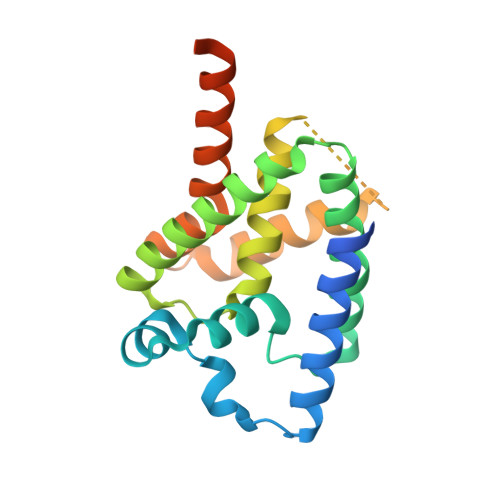
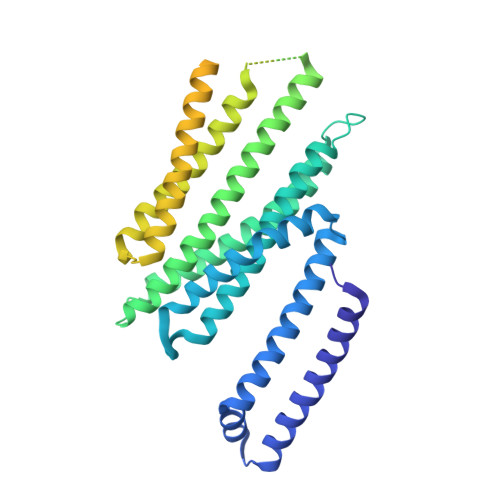
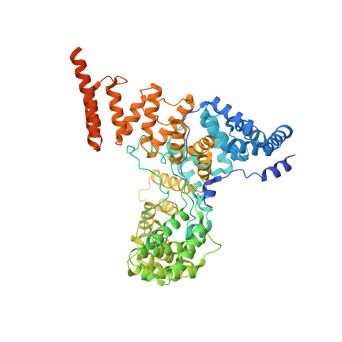
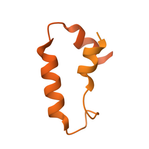
Structural basis of ER-associated protein degradation mediated by the Hrd1 ubiquitin ligase complex.

Wu, X.Siggel, M.Ovchinnikov, S.Mi, W.Svetlov, V.Nudler, E.Liao, M.Hummer, G.Rapoport, T.A.

(2020) Science 368

  • DOI: https://doi.org/10.1126/science.aaz2449
  • Primary Citation of Related Structures:  
    6VJY, 6VJZ, 6VK0, 6VK1, 6VK3

  • PubMed Abstract: 

    Misfolded luminal endoplasmic reticulum (ER) proteins undergo ER-associated degradation (ERAD-L): They are retrotranslocated into the cytosol, polyubiquitinated, and degraded by the proteasome. ERAD-L is mediated by the Hrd1 complex (composed of Hrd1, Hrd3, Der1, Usa1, and Yos9), but the mechanism of retrotranslocation remains mysterious. Here, we report a structure of the active Hrd1 complex, as determined by cryo-electron microscopy analysis of two subcomplexes. Hrd3 and Yos9 jointly create a luminal binding site that recognizes glycosylated substrates. Hrd1 and the rhomboid-like Der1 protein form two "half-channels" with cytosolic and luminal cavities, respectively, and lateral gates facing one another in a thinned membrane region. These structures, along with crosslinking and molecular dynamics simulation results, suggest how a polypeptide loop of an ERAD-L substrate moves through the ER membrane.


  • Organizational Affiliation

    Howard Hughes Medical Institute and Department of Cell Biology, Harvard Medical School, Boston, MA 02115, USA.


Macromolecules
Find similar proteins by:  (by identity cutoff)  |  3D Structure
Entity ID: 1
MoleculeChains Sequence LengthOrganismDetailsImage
Degradation in the endoplasmic reticulum protein 1A [auth C]211Saccharomyces cerevisiaeMutation(s): 0 
Gene Names: DER1
Membrane Entity: Yes 
UniProt
Find proteins for P38307 (Saccharomyces cerevisiae (strain ATCC 204508 / S288c))
Explore P38307 
Go to UniProtKB:  P38307
Entity Groups  
Sequence Clusters30% Identity50% Identity70% Identity90% Identity95% Identity100% Identity
UniProt GroupP38307
Sequence Annotations
Expand
  • Reference Sequence
Find similar proteins by:  (by identity cutoff)  |  3D Structure
Entity ID: 2
MoleculeChains Sequence LengthOrganismDetailsImage
ERAD-associated E3 ubiquitin-protein ligase HRD1480Saccharomyces cerevisiaeMutation(s): 0 
Gene Names: HRD1DER3
EC: 2.3.2.27
Membrane Entity: Yes 
UniProt
Find proteins for Q08109 (Saccharomyces cerevisiae (strain ATCC 204508 / S288c))
Explore Q08109 
Go to UniProtKB:  Q08109
Entity Groups  
Sequence Clusters30% Identity50% Identity70% Identity90% Identity95% Identity100% Identity
UniProt GroupQ08109
Sequence Annotations
Expand
  • Reference Sequence
Find similar proteins by:  (by identity cutoff)  |  3D Structure
Entity ID: 3
MoleculeChains Sequence LengthOrganismDetailsImage
ERAD-associated E3 ubiquitin-protein ligase component HRD3C [auth A]767Saccharomyces cerevisiaeMutation(s): 0 
Gene Names: HRD3
Membrane Entity: Yes 
UniProt
Find proteins for Q05787 (Saccharomyces cerevisiae (strain ATCC 204508 / S288c))
Explore Q05787 
Go to UniProtKB:  Q05787
Entity Groups  
Sequence Clusters30% Identity50% Identity70% Identity90% Identity95% Identity100% Identity
UniProt GroupQ05787
Sequence Annotations
Expand
  • Reference Sequence
Find similar proteins by:  (by identity cutoff)  |  3D Structure
Entity ID: 4
MoleculeChains Sequence LengthOrganismDetailsImage
U1 SNP1-associating protein 1339Saccharomyces cerevisiaeMutation(s): 0 
Gene Names: USA1
Membrane Entity: Yes 
UniProt
Find proteins for Q03714 (Saccharomyces cerevisiae (strain ATCC 204508 / S288c))
Explore Q03714 
Go to UniProtKB:  Q03714
Entity Groups  
Sequence Clusters30% Identity50% Identity70% Identity90% Identity95% Identity100% Identity
UniProt GroupQ03714
Sequence Annotations
Expand
  • Reference Sequence
Experimental Data & Validation

Experimental Data

  • Method: ELECTRON MICROSCOPY
  • Resolution: 4.30 Å
  • Aggregation State: PARTICLE 
  • Reconstruction Method: SINGLE PARTICLE 
EM Software:
TaskSoftware PackageVersion
RECONSTRUCTIONRELION3.0
MODEL REFINEMENTPHENIX

Structure Validation

View Full Validation Report


Entry History & Funding Information

Deposition Data


Funding OrganizationLocationGrant Number
National Institutes of Health/National Institute of General Medical Sciences (NIH/NIGMS)United StatesR01GM052586

Revision History  (Full details and data files)

  • Version 1.0: 2020-04-29
    Type: Initial release
  • Version 1.1: 2020-05-06
    Changes: Database references
  • Version 1.2: 2024-03-06
    Changes: Data collection, Database references