6VGV

Crystal structure of VidaL intein


Experimental Data Snapshot

  • Method: X-RAY DIFFRACTION
  • Resolution: 1.65 Å
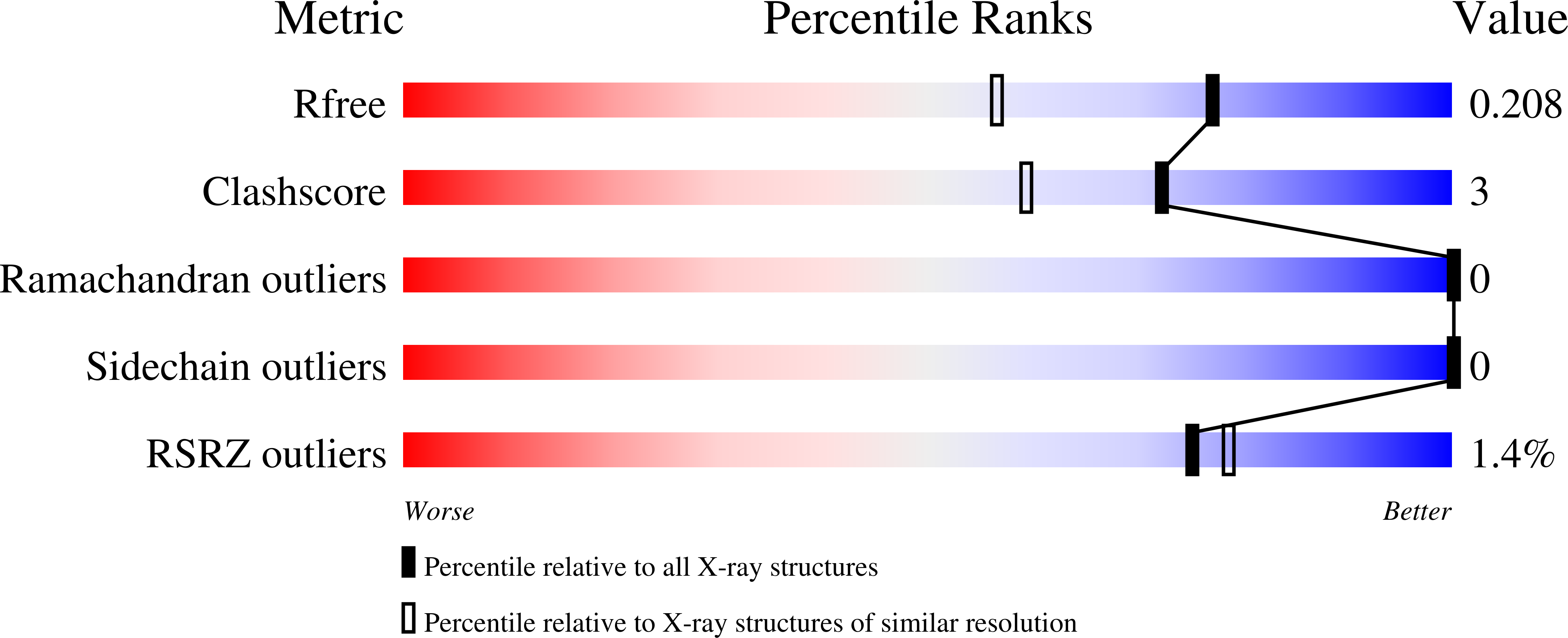
  • R-Value Free: 0.209 
  • R-Value Work: 0.163 
  • R-Value Observed: 0.166 

wwPDB Validation   3D Report Full Report


This is version 1.4 of the entry. See complete history


Literature

Live-cell protein engineering with an ultra-short split intein.

Burton, A.J.Haugbro, M.Parisi, E.Muir, T.W.

(2020) Proc Natl Acad Sci U S A 117: 12041-12049

  • DOI: https://doi.org/10.1073/pnas.2003613117
  • Primary Citation of Related Structures:  
    6VGV, 6VGW

  • PubMed Abstract: 

    Split inteins are privileged molecular scaffolds for the chemical modification of proteins. Though efficient for in vitro applications, these polypeptide ligases have not been utilized for the semisynthesis of proteins in live cells. Here, we biochemically and structurally characterize the naturally split intein VidaL. We show that this split intein, which features the shortest known N-terminal fragment, supports rapid and efficient protein trans -splicing under a range of conditions, enabling semisynthesis of modified proteins both in vitro and in mammalian cells. The utility of this protein engineering system is illustrated through the traceless assembly of multidomain proteins whose biophysical properties render them incompatible with a single expression system, as well as by the semisynthesis of dual posttranslationally modified histone proteins in live cells. We also exploit the domain swapping function of VidaL to effect simultaneous modification and translocation of the nuclear protein HP1α in live cells. Collectively, our studies highlight the VidaL system as a tool for the precise chemical modification of cellular proteins with spatial and temporal control.


  • Organizational Affiliation

    Department of Chemistry, Frick Chemistry Laboratory, Princeton University, Princeton, NJ 08544.


Macromolecules
Find similar proteins by:  (by identity cutoff)  |  3D Structure
Entity ID: 1
MoleculeChains Sequence LengthOrganismDetailsImage
VidaL145synthetic constructMutation(s): 0 
Entity Groups  
Sequence Clusters30% Identity50% Identity70% Identity90% Identity95% Identity100% Identity
Sequence Annotations
Expand
  • Reference Sequence
Experimental Data & Validation

Experimental Data

  • Method: X-RAY DIFFRACTION
  • Resolution: 1.65 Å
  • R-Value Free: 0.209 
  • R-Value Work: 0.163 
  • R-Value Observed: 0.166 
  • Space Group: C 1 2 1
Unit Cell:
Length ( Å )Angle ( ˚ )
a = 100.122α = 90
b = 40.827β = 103.452
c = 36.188γ = 90
Software Package:
Software NamePurpose
PHENIXrefinement
DENZOdata reduction
HKL-3000data scaling
PHASERphasing

Structure Validation

View Full Validation Report


Entry History & Funding Information

Deposition Data


Funding OrganizationLocationGrant Number
National Institutes of Health/National Institute of General Medical Sciences (NIH/NIGMS)United StatesR37-GM086868

Revision History  (Full details and data files)

  • Version 1.0: 2020-05-27
    Type: Initial release
  • Version 1.1: 2020-06-03
    Changes: Database references
  • Version 1.2: 2020-06-17
    Changes: Database references
  • Version 1.3: 2024-03-06
    Changes: Advisory, Data collection, Database references
  • Version 1.4: 2024-04-03
    Changes: Refinement description