6VEC

Cryo-EM structure of F-actin/Plastin2-ABD2 complex


Experimental Data Snapshot

  • Method: ELECTRON MICROSCOPY
  • Resolution: 3.90 Å
  • Aggregation State: FILAMENT 
  • Reconstruction Method: HELICAL 

wwPDB Validation   3D Report Full Report


This is version 1.2 of the entry. See complete history


Literature

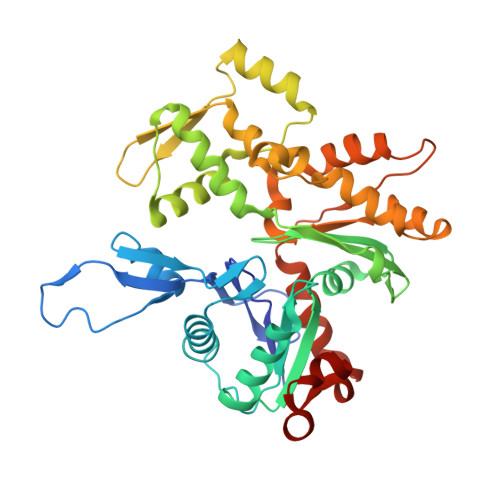
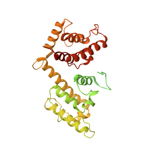
Osteogenesis imperfecta mutations in plastin 3 lead to impaired calcium regulation of actin bundling.

Schwebach, C.L.Kudryashova, E.Zheng, W.Orchard, M.Smith, H.Runyan, L.A.Egelman, E.H.Kudryashov, D.S.

(2020) Bone Res 8: 21-21

  • DOI: https://doi.org/10.1038/s41413-020-0095-2
  • Primary Citation of Related Structures:  
    6VEC

  • PubMed Abstract: 

    Mutations in actin-bundling protein plastin 3 (PLS3) emerged as a cause of congenital osteoporosis, but neither the role of PLS3 in bone development nor the mechanisms underlying PLS3-dependent osteoporosis are understood. Of the over 20 identified osteoporosis-linked PLS3 mutations, we investigated all five that are expected to produce full-length protein. One of the mutations distorted an actin-binding loop in the second actin-binding domain of PLS3 and abolished F-actin bundling as revealed by cryo-EM reconstruction and protein interaction assays. Surprisingly, the remaining four mutants fully retained F-actin bundling ability. However, they displayed defects in Ca 2+ sensitivity: two of the mutants lost the ability to be inhibited by Ca 2+ , while the other two became hypersensitive to Ca 2+ . Each group of the mutants with similar biochemical properties showed highly characteristic cellular behavior. Wild-type PLS3 was distributed between lamellipodia and focal adhesions. In striking contrast, the Ca 2+ -hyposensitive mutants were not found at the leading edge but localized exclusively at focal adhesions/stress fibers, which displayed reinforced morphology. Consistently, the Ca 2+ -hypersensitive PLS3 mutants were restricted to lamellipodia, while chelation of Ca 2+ caused their redistribution to focal adhesions. Finally, the bundling-deficient mutant failed to co-localize with any F-actin structures in cells despite a preserved F-actin binding through a non-mutation-bearing actin-binding domain. Our findings revealed that severe osteoporosis can be caused by a mutational disruption of the Ca 2+ -controlled PLS3's cycling between adhesion complexes and the leading edge. Integration of the structural, biochemical, and cell biology insights enabled us to propose a molecular mechanism of plastin activity regulation by Ca 2+ .


  • Organizational Affiliation

    Department of Chemistry and Biochemistry, The Ohio State University, Columbus, OH 43210 USA.


Macromolecules
Find similar proteins by:  (by identity cutoff)  |  3D Structure
Entity ID: 1
MoleculeChains Sequence LengthOrganismDetailsImage
Actin, alpha skeletal muscle
A, B, C, D, E
A, B, C, D, E, F, G, H, I, J, K
377Oryctolagus cuniculusMutation(s): 0 
UniProt
Find proteins for P68135 (Oryctolagus cuniculus)
Explore P68135 
Go to UniProtKB:  P68135
Entity Groups  
Sequence Clusters30% Identity50% Identity70% Identity90% Identity95% Identity100% Identity
UniProt GroupP68135
Sequence Annotations
Expand
  • Reference Sequence
Find similar proteins by:  (by identity cutoff)  |  3D Structure
Entity ID: 2
MoleculeChains Sequence LengthOrganismDetailsImage
LCP1422Homo sapiensMutation(s): 0 
Gene Names: HEL-S-37
UniProt & NIH Common Fund Data Resources
Find proteins for P13796 (Homo sapiens)
Explore P13796 
Go to UniProtKB:  P13796
PHAROS:  P13796
GTEx:  ENSG00000136167 
Entity Groups  
Sequence Clusters30% Identity50% Identity70% Identity90% Identity95% Identity100% Identity
UniProt GroupP13796
Sequence Annotations
Expand
  • Reference Sequence
Small Molecules
Ligands 2 Unique
IDChains Name / Formula / InChI Key2D Diagram3D Interactions
ADP
Query on ADP

Download Ideal Coordinates CCD File 
AA [auth C]
CA [auth D]
EA [auth E]
GA [auth F]
IA [auth G]
AA [auth C],
CA [auth D],
EA [auth E],
GA [auth F],
IA [auth G],
KA [auth H],
MA [auth I],
OA [auth J],
QA [auth K],
W [auth A],
Y [auth B]
ADENOSINE-5'-DIPHOSPHATE
C10 H15 N5 O10 P2
XTWYTFMLZFPYCI-KQYNXXCUSA-N
MG
Query on MG

Download Ideal Coordinates CCD File 
BA [auth C]
DA [auth D]
FA [auth E]
HA [auth F]
JA [auth G]
BA [auth C],
DA [auth D],
FA [auth E],
HA [auth F],
JA [auth G],
LA [auth H],
NA [auth I],
PA [auth J],
RA [auth K],
X [auth A],
Z [auth B]
MAGNESIUM ION
Mg
JLVVSXFLKOJNIY-UHFFFAOYSA-N
Experimental Data & Validation

Experimental Data

  • Method: ELECTRON MICROSCOPY
  • Resolution: 3.90 Å
  • Aggregation State: FILAMENT 
  • Reconstruction Method: HELICAL 
EM Software:
TaskSoftware PackageVersion
RECONSTRUCTIONRELION
MODEL REFINEMENTCoot
MODEL REFINEMENTPHENIX

Structure Validation

View Full Validation Report


Entry History & Funding Information

Deposition Data


Funding OrganizationLocationGrant Number
National Institutes of Health/National Institute of General Medical Sciences (NIH/NIGMS)United StatesR35GM122510

Revision History  (Full details and data files)

  • Version 1.0: 2020-12-09
    Type: Initial release
  • Version 1.1: 2020-12-23
    Changes: Database references, Structure summary
  • Version 1.2: 2024-03-06
    Changes: Data collection, Database references, Refinement description