6VD7

Cryo-EM structure of Xenopus tropicalis pannexin 1 channel


Experimental Data Snapshot

  • Method: ELECTRON MICROSCOPY
  • Resolution: 3.02 Å
  • Aggregation State: PARTICLE 
  • Reconstruction Method: SINGLE PARTICLE 

wwPDB Validation   3D Report Full Report


This is version 1.1 of the entry. See complete history


Literature

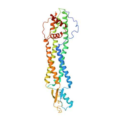
The Cryo-EM structure of pannexin 1 reveals unique motifs for ion selection and inhibition.

Michalski, K.Syrjanen, J.L.Henze, E.Kumpf, J.Furukawa, H.Kawate, T.

(2020) Elife 9

  • DOI: https://doi.org/10.7554/eLife.54670
  • Primary Citation of Related Structures:  
    6VD7

  • PubMed Abstract: 

    Pannexins are large-pore forming channels responsible for ATP release under a variety of physiological and pathological conditions. Although predicted to share similar membrane topology with other large-pore forming proteins such as connexins, innexins, and LRRC8, pannexins have minimal sequence similarity to these protein families. Here, we present the cryo-EM structure of a frog pannexin 1 (Panx1) channel at 3.0 Å. We find that Panx1 protomers harbor four transmembrane helices similar in arrangement to other large-pore forming proteins but assemble as a heptameric channel with a unique constriction formed by Trp74 in the first extracellular loop. Mutating Trp74 or the nearby Arg75 disrupt ion selectivity, whereas altering residues in the hydrophobic groove formed by the two extracellular loops abrogates channel inhibition by carbenoxolone. Our structural and functional study establishes the extracellular loops as important structural motifs for ion selectivity and channel inhibition in Panx1.


  • Organizational Affiliation

    Department of Molecular Medicine, Cornell University, Ithaca, United States.


Macromolecules
Find similar proteins by:  (by identity cutoff)  |  3D Structure
Entity ID: 1
MoleculeChains Sequence LengthOrganismDetailsImage
Pannexin
A, B, C, D, E
A, B, C, D, E, F, G
344Xenopus tropicalisMutation(s): 0 
Gene Names: panx1igdcc4LOC100170473nell1PANX
Membrane Entity: Yes 
UniProt
Find proteins for B3DLA5 (Xenopus tropicalis)
Explore B3DLA5 
Go to UniProtKB:  B3DLA5
Entity Groups  
Sequence Clusters30% Identity50% Identity70% Identity90% Identity95% Identity100% Identity
UniProt GroupB3DLA5
Sequence Annotations
Expand
  • Reference Sequence
Experimental Data & Validation

Experimental Data

  • Method: ELECTRON MICROSCOPY
  • Resolution: 3.02 Å
  • Aggregation State: PARTICLE 
  • Reconstruction Method: SINGLE PARTICLE 
EM Software:
TaskSoftware PackageVersion
RECONSTRUCTIONcisTEM
MODEL REFINEMENTPHENIX

Structure Validation

View Full Validation Report


Entry History & Funding Information

Deposition Data


Funding OrganizationLocationGrant Number
National Institutes of Health/National Institute of General Medical Sciences (NIH/NIGMS)United States--

Revision History  (Full details and data files)

  • Version 1.0: 2020-02-26
    Type: Initial release
  • Version 1.1: 2020-04-15
    Changes: Database references