6V67

Apo Structure of the De Novo PD-1 Binding Miniprotein GR918.2


Experimental Data Snapshot

  • Method: X-RAY DIFFRACTION
  • Resolution: 1.07 Å
  • R-Value Free: 0.150 
  • R-Value Work: 0.145 
  • R-Value Observed: 0.146 

wwPDB Validation   3D Report Full Report


This is version 1.2 of the entry. See complete history


Literature

Computational design of a synthetic PD-1 agonist.

Bryan, C.M.Rocklin, G.J.Bick, M.J.Ford, A.Majri-Morrison, S.Kroll, A.V.Miller, C.J.Carter, L.Goreshnik, I.Kang, A.DiMaio, F.Tarbell, K.V.Baker, D.

(2021) Proc Natl Acad Sci U S A 118

  • DOI: https://doi.org/10.1073/pnas.2102164118
  • Primary Citation of Related Structures:  
    6V67

  • PubMed Abstract: 

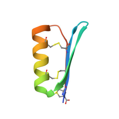
    Programmed cell death protein-1 (PD-1) expressed on activated T cells inhibits T cell function and proliferation to prevent an excessive immune response, and disease can result if this delicate balance is shifted in either direction. Tumor cells often take advantage of this pathway by overexpressing the PD-1 ligand PD-L1 to evade destruction by the immune system. Alternatively, if there is a decrease in function of the PD-1 pathway, unchecked activation of the immune system and autoimmunity can result. Using a combination of computation and experiment, we designed a hyperstable 40-residue miniprotein, PD-MP1, that specifically binds murine and human PD-1 at the PD-L1 interface with a K d of ∼100 nM. The apo crystal structure shows that the binder folds as designed with a backbone RMSD of 1.3 Å to the design model. Trimerization of PD-MP1 resulted in a PD-1 agonist that strongly inhibits murine T cell activation. This small, hyperstable PD-1 binding protein was computationally designed with an all-beta interface, and the trimeric agonist could contribute to treatments for autoimmune and inflammatory diseases.


  • Organizational Affiliation

    Department of Biochemistry, University of Washington, Seattle, WA 98195; cassie.bryan@gmail.com.


Macromolecules
Find similar proteins by:  (by identity cutoff)  |  3D Structure
Entity ID: 1
MoleculeChains Sequence LengthOrganismDetailsImage
PD-1 Binding Miniprotein GR918.2
A, B
43synthetic constructMutation(s): 0 
Entity Groups  
Sequence Clusters30% Identity50% Identity70% Identity90% Identity95% Identity100% Identity
Sequence Annotations
Expand
  • Reference Sequence
Experimental Data & Validation

Experimental Data

  • Method: X-RAY DIFFRACTION
  • Resolution: 1.07 Å
  • R-Value Free: 0.150 
  • R-Value Work: 0.145 
  • R-Value Observed: 0.146 
  • Space Group: C 1 2 1
Unit Cell:
Length ( Å )Angle ( ˚ )
a = 50.849α = 90
b = 26.744β = 99.67
c = 51.537γ = 90
Software Package:
Software NamePurpose
XDSdata reduction
Aimlessdata scaling
PHENIXrefinement
PDB_EXTRACTdata extraction
PHASERphasing

Structure Validation

View Full Validation Report


Entry History & Funding Information

Deposition Data


Funding OrganizationLocationGrant Number
National Institutes of Health/National Institute of General Medical Sciences (NIH/NIGMS)United States5U01GM094665
National Institutes of Health/National Institute of General Medical Sciences (NIH/NIGMS)United StatesGM123089

Revision History  (Full details and data files)

  • Version 1.0: 2020-12-09
    Type: Initial release
  • Version 1.1: 2021-12-29
    Changes: Database references
  • Version 1.2: 2024-04-03
    Changes: Data collection, Refinement description