6V04

DynU16 crystal structure, a putative protein in the dynemicin biosynthetic locus


Experimental Data Snapshot

  • Method: X-RAY DIFFRACTION
  • Resolution: 1.50 Å
  • R-Value Free: 0.188 
  • R-Value Work: 0.162 
  • R-Value Observed: 0.163 

wwPDB Validation   3D Report Full Report


This is version 2.2 of the entry. See complete history


Literature

Structural characterization of DynU16, a START/Bet v1-like protein involved in dynemicin biosynthesis.

Alvarado, S.K.Miller, M.D.Bhardwaj, M.Thorson, J.S.Van Lanen, S.G.Phillips Jr., G.N.

(2021) Acta Crystallogr F Struct Biol Commun 77: 328-333

  • DOI: https://doi.org/10.1107/S2053230X21008943
  • Primary Citation of Related Structures:  
    6V04

  • PubMed Abstract: 

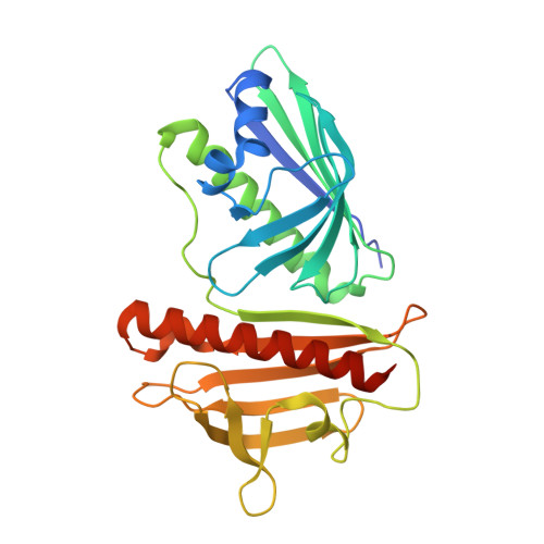
    The 1.5 Å resolution crystal structure of DynU16, a protein identified in the dynemicin-biosynthetic gene cluster, is reported. The structure adopts a di-domain helix-grip fold with a uniquely positioned open cavity connecting the domains. The elongated dimensions of the cavity appear to be compatible with the geometry of a linear polyene, suggesting the involvement of DynU16 in the upstream steps of dynemicin biosynthesis.


  • Organizational Affiliation

    Department of BioSciences, Rice University, 6100 Main Street, Houston, TX 77005, USA.


Macromolecules
Find similar proteins by:  (by identity cutoff)  |  3D Structure
Entity ID: 1
MoleculeChains Sequence LengthOrganismDetailsImage
Uncharacterized SRPBCC domain-containing protein284Micromonospora chersinaMutation(s): 0 
Gene Names: GA0070603_4194
UniProt
Find proteins for B2BM46 (Micromonospora chersina)
Explore B2BM46 
Go to UniProtKB:  B2BM46
Entity Groups  
Sequence Clusters30% Identity50% Identity70% Identity90% Identity95% Identity100% Identity
UniProt GroupB2BM46
Sequence Annotations
Expand
  • Reference Sequence
Experimental Data & Validation

Experimental Data

  • Method: X-RAY DIFFRACTION
  • Resolution: 1.50 Å
  • R-Value Free: 0.188 
  • R-Value Work: 0.162 
  • R-Value Observed: 0.163 
  • Space Group: P 31 2 1
Unit Cell:
Length ( Å )Angle ( ˚ )
a = 73.171α = 90
b = 73.171β = 90
c = 123.944γ = 120
Software Package:
Software NamePurpose
PDB_EXTRACTdata extraction
PHENIXrefinement
SHELXphasing
HKL2Mapphasing
XDSdata scaling
XDSdata reduction
xia2data reduction

Structure Validation

View Full Validation Report


Entry History & Funding Information

Deposition Data


Funding OrganizationLocationGrant Number
National Institutes of Health/National Institute of General Medical Sciences (NIH/NIGMS)United StatesR01 GM115261
National Institutes of Health/National Cancer Institute (NIH/NCI)United StatesR01-CA217255
National Science Foundation (NSF, United States)United StatesSTC 1231306

Revision History  (Full details and data files)

  • Version 1.0: 2020-11-18
    Type: Initial release
  • Version 2.0: 2021-04-28
    Changes: Advisory, Atomic model, Data collection, Database references, Derived calculations, Polymer sequence, Source and taxonomy, Structure summary
  • Version 2.1: 2021-09-15
    Changes: Database references, Structure summary
  • Version 2.2: 2022-04-06
    Changes: Database references