6UUJ

Structure of PE5-PPE4-EspG3 complex from the type VII (ESX-3) secretion system, space group P212121


Experimental Data Snapshot

  • Method: X-RAY DIFFRACTION
  • Resolution: 3.00 Å
  • R-Value Free: 0.301 
  • R-Value Work: 0.266 
  • R-Value Observed: 0.268 

wwPDB Validation   3D Report Full Report

Currently 6UUJ does not have a validation slider image.


This is version 1.2 of the entry. See complete history


Literature

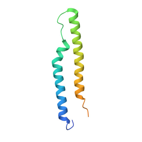
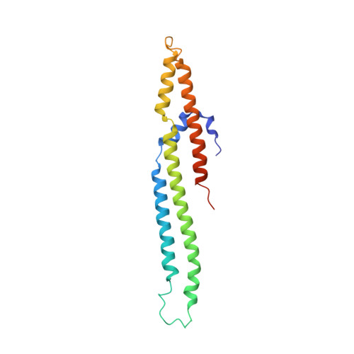
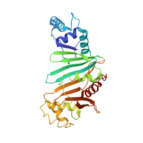
PE5-PPE4-EspG3heterotrimer structure from mycobacterial ESX-3 secretion system gives insight into cognate substrate recognition by ESX systems.

Williamson, Z.A.Chaton, C.T.Ciocca, W.A.Korotkova, N.Korotkov, K.V.

(2020) J Biol Chem 295: 12706-12715

  • DOI: https://doi.org/10.1074/jbc.RA120.012698
  • Primary Citation of Related Structures:  
    6UUJ, 6VHR

  • PubMed Abstract: 

    Mycobacterium tuberculosis has evolved numerous type VII secretion (ESX) systems to secrete multiple factors important for both growth and virulence across their cell envelope. ESX-1, ESX-3, and ESX-5 systems have been shown to each secrete a distinct set of substrates, including PE and PPE families of proteins, named for conserved Pro-Glu and Pro-Pro-Glu motifs in their N termini. Proper secretion of the PE-PPE proteins requires the presence of EspG, with each system encoding its own unique copy. There is no cross-talk between any of the ESX systems, and how each EspG recognizes its subset of PE-PPE proteins is currently unknown. The only current structural characterization of PE-PPE-EspG heterotrimers is from the ESX-5 system. Here we present the crystal structure of the PE5 mt -PPE4 mt -EspG 3mm heterotrimer from the ESX-3 system. Our heterotrimer reveals that EspG 3mm interacts exclusively with PPE4 mt in a similar manner to EspG 5 , shielding the hydrophobic tip of PPE4 mt from solvent. The C-terminal helical domain of EspG 3mm is dynamic, alternating between "open" and "closed" forms, and this movement is likely functionally relevant in the unloading of PE-PPE heterodimers at the secretion machinery. In contrast to the previously solved ESX-5 heterotrimers, the PE-PPE heterodimer of our ESX-3 heterotrimer is interacting with its chaperone at a drastically different angle and presents different faces of the PPE protein to the chaperone. We conclude that the PPE-EspG interface from each ESX system has a unique shape complementarity that allows each EspG to discriminate among noncognate PE-PPE pairs.


  • Organizational Affiliation

    Department of Molecular & Cellular Biochemistry and the Center for Structural Biology, University of Kentucky, Lexington, Kentucky, USA.


Macromolecules
Find similar proteins by:  (by identity cutoff)  |  3D Structure
Entity ID: 1
MoleculeChains Sequence LengthOrganismDetailsImage
PE family immunomodulator PE5
A, D, G, J
100Mycobacterium tuberculosis H37RvMutation(s): 0 
Gene Names: PE5Rv0285LH57_01560
UniProt
Find proteins for L7N695 (Mycobacterium tuberculosis (strain ATCC 25618 / H37Rv))
Explore L7N695 
Go to UniProtKB:  L7N695
Entity Groups  
Sequence Clusters30% Identity50% Identity70% Identity90% Identity95% Identity100% Identity
UniProt GroupL7N695
Sequence Annotations
Expand
  • Reference Sequence
Find similar proteins by:  (by identity cutoff)  |  3D Structure
Entity ID: 2
MoleculeChains Sequence LengthOrganismDetailsImage
PPE family protein PPE4
B, E, H, K
178Mycobacterium tuberculosis H37RvMutation(s): 0 
Gene Names: PPE4Rv0286
UniProt
Find proteins for P9WI43 (Mycobacterium tuberculosis (strain ATCC 25618 / H37Rv))
Explore P9WI43 
Go to UniProtKB:  P9WI43
Entity Groups  
Sequence Clusters30% Identity50% Identity70% Identity90% Identity95% Identity100% Identity
UniProt GroupP9WI43
Sequence Annotations
Expand
  • Reference Sequence
Find similar proteins by:  (by identity cutoff)  |  3D Structure
Entity ID: 3
MoleculeChains Sequence LengthOrganismDetailsImage
ESX-3 secretion-associated protein EspG3
C, F, I, L
288Mycobacterium marinum MMutation(s): 0 
Gene Names: MMAR_0548
UniProt
Find proteins for B2HNX0 (Mycobacterium marinum (strain ATCC BAA-535 / M))
Explore B2HNX0 
Go to UniProtKB:  B2HNX0
Entity Groups  
Sequence Clusters30% Identity50% Identity70% Identity90% Identity95% Identity100% Identity
UniProt GroupB2HNX0
Sequence Annotations
Expand
  • Reference Sequence
Experimental Data & Validation

Experimental Data

  • Method: X-RAY DIFFRACTION
  • Resolution: 3.00 Å
  • R-Value Free: 0.301 
  • R-Value Work: 0.266 
  • R-Value Observed: 0.268 
  • Space Group: P 21 21 21
Unit Cell:
Length ( Å )Angle ( ˚ )
a = 72.265α = 90
b = 158.63β = 90
c = 209.314γ = 90
Software Package:
Software NamePurpose
XSCALEdata scaling
PHENIXrefinement
PDB_EXTRACTdata extraction
XDSdata reduction
PHASERphasing

Structure Validation

View Full Validation Report

Currently 6UUJ does not have a validation slider image.


Entry History & Funding Information

Deposition Data


Funding OrganizationLocationGrant Number
National Institutes of Health/National Institute Of Allergy and Infectious Diseases (NIH/NIAID)United StatesR01AI119022
National Institutes of Health/National Institute of General Medical Sciences (NIH/NIGMS)United StatesP20GM103486
National Institutes of Health/National Institute of General Medical Sciences (NIH/NIGMS)United StatesP30GM110787

Revision History  (Full details and data files)

  • Version 1.0: 2020-01-29
    Type: Initial release
  • Version 1.1: 2020-09-16
    Changes: Database references
  • Version 1.2: 2023-10-11
    Changes: Data collection, Database references, Refinement description