6UUG

Structure of methanesulfinate monooxygenase MsuC from Pseudomonas fluorescens at 1.69 angstrom resolution


Experimental Data Snapshot

  • Method: X-RAY DIFFRACTION
  • Resolution: 1.69 Å
  • R-Value Free: 0.208 
  • R-Value Work: 0.185 
  • R-Value Observed: 0.186 

wwPDB Validation   3D Report Full Report

Currently 6UUG does not have a validation slider image.


This is version 1.4 of the entry. See complete history


Literature

Structure and function of the two-component flavin-dependent methanesulfinate monooxygenase within bacterial sulfur assimilation.

Soule, J.Gnann, A.D.Gonzalez, R.Parker, M.J.McKenna, K.C.Nguyen, S.V.Phan, N.T.Wicht, D.K.Dowling, D.P.

(2020) Biochem Biophys Res Commun 522: 107-112

  • DOI: https://doi.org/10.1016/j.bbrc.2019.11.008
  • Primary Citation of Related Structures:  
    6UUG

  • PubMed Abstract: 

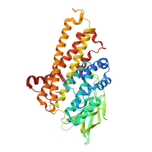
    Methyl sulfur compounds are a rich source of environmental sulfur for microorganisms, but their use requires redox systems. The bacterial sfn and msu operons contain two-component flavin-dependent monooxygenases for dimethylsulfone (DMSO 2 ) assimilation: SfnG converts DMSO 2 to methanesulfinate (MSI - ), and MsuD converts methanesulfonate (MS - ) to sulfite. However, the enzymatic oxidation of MSI - to MS - has not been demonstrated, and the function of the last enzyme of the msu operon (MsuC) is unresolved. We employed crystallographic and biochemical studies to identify the function of MsuC from Pseudomonas fluorescens. The crystal structure of MsuC adopts the acyl-CoA dehydrogenase fold with putative binding sites for flavin and MSI - , and functional assays of MsuC in the presence of its oxidoreductase MsuE, FMN, and NADH confirm the enzymatic generation of MS - . These studies reveal that MsuC converts MSI - to MS - in sulfite biosynthesis from DMSO 2 .


  • Organizational Affiliation

    Department of Chemistry, University of Massachusetts Boston, Boston, MA, 02125, USA.


Macromolecules
Find similar proteins by:  (by identity cutoff)  |  3D Structure
Entity ID: 1
MoleculeChains Sequence LengthOrganismDetailsImage
Putative dehydrogenase
A, B
415Pseudomonas fluorescens Pf0-1Mutation(s): 0 
Gene Names: Pfl01_3917
UniProt
Find proteins for Q3K9A0 (Pseudomonas fluorescens (strain Pf0-1))
Explore Q3K9A0 
Go to UniProtKB:  Q3K9A0
Entity Groups  
Sequence Clusters30% Identity50% Identity70% Identity90% Identity95% Identity100% Identity
UniProt GroupQ3K9A0
Sequence Annotations
Expand
  • Reference Sequence
Experimental Data & Validation

Experimental Data

  • Method: X-RAY DIFFRACTION
  • Resolution: 1.69 Å
  • R-Value Free: 0.208 
  • R-Value Work: 0.185 
  • R-Value Observed: 0.186 
  • Space Group: P 21 21 2
Unit Cell:
Length ( Å )Angle ( ˚ )
a = 81.187α = 90
b = 161.582β = 90
c = 62.045γ = 90
Software Package:
Software NamePurpose
PHENIXrefinement
XDSdata reduction
XDSdata scaling
PHASERphasing
Cootmodel building

Structure Validation

View Full Validation Report

Currently 6UUG does not have a validation slider image.


Entry History & Funding Information

Deposition Data


Funding OrganizationLocationGrant Number
National Science Foundation (NSF, United States)United States1807480

Revision History  (Full details and data files)

  • Version 1.0: 2019-12-04
    Type: Initial release
  • Version 1.1: 2020-01-08
    Changes: Author supporting evidence
  • Version 1.2: 2020-01-22
    Changes: Database references
  • Version 1.3: 2020-05-06
    Changes: Structure summary
  • Version 1.4: 2023-10-11
    Changes: Data collection, Database references, Refinement description