6UQR

Complex of IgE and Ligelizumab

  • Classification: IMMUNE SYSTEM
  • Organism(s): Homo sapiens
  • Expression System: Homo sapiens
  • Mutation(s): No 

  • Deposited: 2019-10-21 Released: 2019-12-04 
  • Deposition Author(s): Tarchevskaya, S.S., Kleinboelting, S., Jardetzky, T.S.
  • Funding Organization(s): National Institutes of Health/National Institute Of Allergy and Infectious Diseases (NIH/NIAID), National Institutes of Health/National Heart, Lung, and Blood Institute (NIH/NHLBI)

Experimental Data Snapshot

  • Method: X-RAY DIFFRACTION
  • Resolution: 3.65 Å
  • R-Value Free: 0.295 
  • R-Value Work: 0.290 
  • R-Value Observed: 0.291 

wwPDB Validation   3D Report Full Report

Currently 6UQR does not have a validation slider image.


This is version 2.1 of the entry. See complete history


Literature

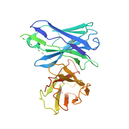
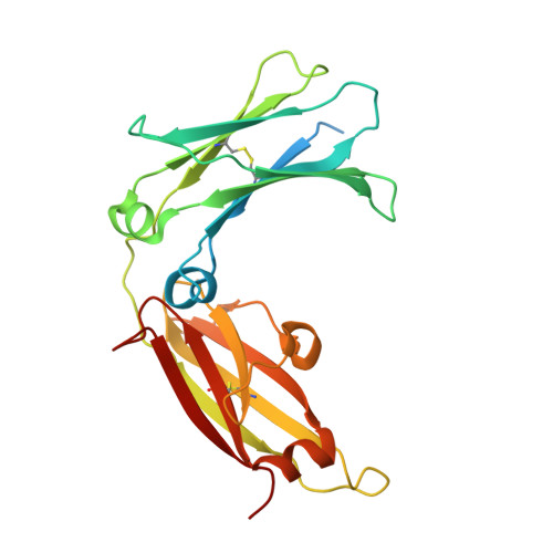
The mechanistic and functional profile of the therapeutic anti-IgE antibody ligelizumab differs from omalizumab.

Gasser, P.Tarchevskaya, S.S.Guntern, P.Brigger, D.Ruppli, R.Zbaren, N.Kleinboelting, S.Heusser, C.Jardetzky, T.S.Eggel, A.

(2020) Nat Commun 11: 165-165

  • DOI: https://doi.org/10.1038/s41467-019-13815-w
  • Primary Citation of Related Structures:  
    6UQR

  • PubMed Abstract: 

    Targeting of immunoglobulin E (IgE) represents an interesting approach for the treatment of allergic disorders. A high-affinity monoclonal anti-IgE antibody, ligelizumab, has recently been developed to overcome some of the limitations associated with the clinical use of the therapeutic anti-IgE antibody, omalizumab. Here, we determine the molecular binding profile and functional modes-of-action of ligelizumab. We solve the crystal structure of ligelizumab bound to IgE, and report epitope differences between ligelizumab and omalizumab that contribute to their qualitatively distinct IgE-receptor inhibition profiles. While ligelizumab shows superior inhibition of IgE binding to FcεRI, basophil activation, IgE production by B cells and passive systemic anaphylaxis in an in vivo mouse model, ligelizumab is less potent in inhibiting IgE:CD23 interactions than omalizumab. Our data thus provide a structural and mechanistic foundation for understanding the efficient suppression of FcεRI-dependent allergic reactions by ligelizumab in vitro as well as in vivo.


  • Organizational Affiliation

    Department of BioMedical Research, University of Bern, Bern, Switzerland.


Macromolecules
Find similar proteins by:  (by identity cutoff)  |  3D Structure
Entity ID: 1
MoleculeChains Sequence LengthOrganismDetailsImage
Ligelizumab
A, C
268Homo sapiensMutation(s): 0 
Entity Groups  
Sequence Clusters30% Identity50% Identity70% Identity90% Identity95% Identity100% Identity
Sequence Annotations
Expand
  • Reference Sequence
Find similar proteins by:  (by identity cutoff)  |  3D Structure
Entity ID: 2
MoleculeChains Sequence LengthOrganismDetailsImage
IgE
B, D
247Homo sapiensMutation(s): 0 
Gene Names: IGHE
UniProt & NIH Common Fund Data Resources
Find proteins for P01854 (Homo sapiens)
Explore P01854 
Go to UniProtKB:  P01854
PHAROS:  P01854
GTEx:  ENSG00000211891 
Entity Groups  
Sequence Clusters30% Identity50% Identity70% Identity90% Identity95% Identity100% Identity
UniProt GroupP01854
Sequence Annotations
Expand
  • Reference Sequence
Oligosaccharides

Help

Entity ID: 3
MoleculeChains Length2D Diagram Glycosylation3D Interactions
alpha-D-mannopyranose-(1-3)-[alpha-D-mannopyranose-(1-6)]beta-D-mannopyranose-(1-4)-2-acetamido-2-deoxy-beta-D-glucopyranose-(1-4)-2-acetamido-2-deoxy-beta-D-glucopyranose
E
5N-Glycosylation
Glycosylation Resources
GlyTouCan:  G22768VO
GlyCosmos:  G22768VO
GlyGen:  G22768VO
Entity ID: 4
MoleculeChains Length2D Diagram Glycosylation3D Interactions
alpha-D-mannopyranose-(1-3)-beta-D-mannopyranose-(1-4)-2-acetamido-2-deoxy-beta-D-glucopyranose-(1-4)-2-acetamido-2-deoxy-beta-D-glucopyranose
F
4N-Glycosylation
Glycosylation Resources
GlyTouCan:  G81315DD
GlyCosmos:  G81315DD
GlyGen:  G81315DD
Experimental Data & Validation

Experimental Data

  • Method: X-RAY DIFFRACTION
  • Resolution: 3.65 Å
  • R-Value Free: 0.295 
  • R-Value Work: 0.290 
  • R-Value Observed: 0.291 
  • Space Group: P 21 21 21
Unit Cell:
Length ( Å )Angle ( ˚ )
a = 74.96α = 90
b = 103.18β = 90
c = 124.61γ = 90
Software Package:
Software NamePurpose
PHENIXrefinement
XDSdata reduction
XSCALEdata scaling
PHASERphasing

Structure Validation

View Full Validation Report

Currently 6UQR does not have a validation slider image.


Entry History & Funding Information

Deposition Data


Funding OrganizationLocationGrant Number
National Institutes of Health/National Institute Of Allergy and Infectious Diseases (NIH/NIAID)United StatesR01 AI115469
National Institutes of Health/National Heart, Lung, and Blood Institute (NIH/NHLBI)United StatesR01 HL141493

Revision History  (Full details and data files)

  • Version 1.0: 2019-12-04
    Type: Initial release
  • Version 1.1: 2020-04-01
    Changes: Data collection, Database references
  • Version 2.0: 2020-07-29
    Type: Remediation
    Reason: Carbohydrate remediation
    Changes: Atomic model, Data collection, Derived calculations, Structure summary
  • Version 2.1: 2023-10-11
    Changes: Data collection, Database references, Derived calculations, Refinement description, Structure summary