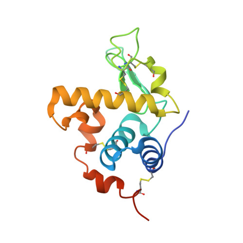
Crystal structure of a C-type lysozyme from Litopenaeus vanamei exhibiting a high binding constant to its chitotriose inhibitor.
Vargas-Requena, C.L., Rodriguez-Romero, A., Garcia-Ramirez, B., Sotelo-Mundo, R.R., Hernandez-Santoyo, A.(2020) Fish Shellfish Immunol 100: 246-255
- PubMed: 32151687
- DOI: https://doi.org/10.1016/j.fsi.2020.03.010
- Primary Citation of Related Structures:
6UKC, 6UL3 - PubMed Abstract:
Although information about invertebrate lysozymes is scarce, these enzymes have been described as components of the innate immune system, functioning as antibacterial proteins. Here we describe the first thermodynamic and structural study of a new C-type lysozyme from a Pacific white shrimp Litopenaeus vannamei (LvL), which has shown high activity against both Gram (+) and Gram (-) bacteria including Vibrio sp. that is one of the most severe pathogens in penaeid shrimp aquaculture. Compared with hen egg-white lysozyme, its sequence harbors a seven-residue insertion from amino acid 97 to 103, and a nine-residue extension at the C-terminus only found in penaeid crustaceans, making this enzyme one of the longest lysozyme reported to date. LvL was crystallized in the presence and absence of chitotriose. The former crystallized as a monomer in space group P6 1 and the latter in P2 1 2 1 2 1 with two monomers in the asymmetric unit. Since the enzyme crystallized at a pH where lysozyme activity is deficient, the ligand could not be observed in the P6 1 structure; therefore, we performed a docking simulation with chitotriose to compare with the hen egg lysozyme crystallized in the presence of the ligand. Remarkably, additional amino acids in LvL caused an increase in the length of α-helix H4 (residues 97-103) that is directly related to ligand recognition. The K a for chitotriose (4.1 × 10 5 M -1 ), as determined by Isothermal Titration Calorimetry, was one order of magnitude higher than those for lysozymes from hen and duck eggs. Our results revealed new interactions of chitiotriose with residues in helix H4.
Organizational Affiliation:
Instituto de Ciencias Biomédicas, Universidad Autónoma de Ciudad Juárez, Cd. Juárez Chihuaha, 32000, Mexico.