6UEB

Structure of Rabies SAD-B19 L-P complex from cryo-EM


Experimental Data Snapshot

  • Method: ELECTRON MICROSCOPY
  • Resolution: 3.30 Å
  • Aggregation State: PARTICLE 
  • Reconstruction Method: SINGLE PARTICLE 

wwPDB Validation   3D Report Full Report


This is version 1.2 of the entry. See complete history


Literature

Structure of a rabies virus polymerase complex from electron cryo-microscopy.

Horwitz, J.A.Jenni, S.Harrison, S.C.Whelan, S.P.J.

(2020) Proc Natl Acad Sci U S A 117: 2099-2107

  • DOI: https://doi.org/10.1073/pnas.1918809117
  • Primary Citation of Related Structures:  
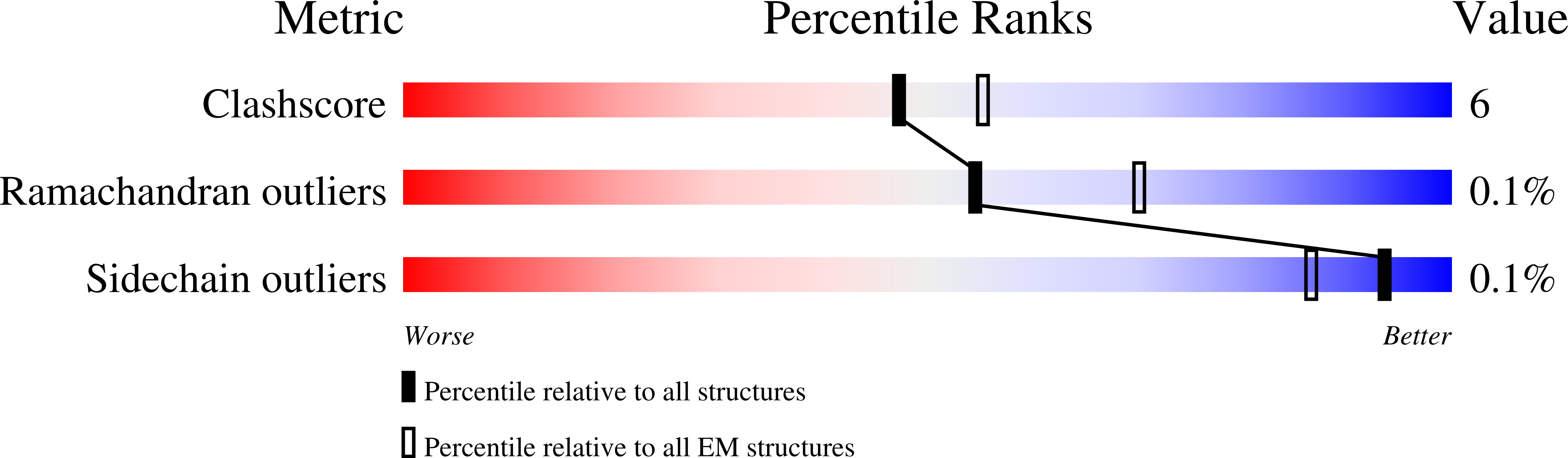
    6UEB

  • PubMed Abstract: 

    Nonsegmented negative-stranded (NNS) RNA viruses, among them the virus that causes rabies (RABV), include many deadly human pathogens. The large polymerase (L) proteins of NNS RNA viruses carry all of the enzymatic functions required for viral messenger RNA (mRNA) transcription and replication: RNA polymerization, mRNA capping, and cap methylation. We describe here a complete structure of RABV L bound with its phosphoprotein cofactor (P), determined by electron cryo-microscopy at 3.3 Å resolution. The complex closely resembles the vesicular stomatitis virus (VSV) L-P, the one other known full-length NNS-RNA L-protein structure, with key local differences (e.g., in L-P interactions). Like the VSV L-P structure, the RABV complex analyzed here represents a preinitiation conformation. Comparison with the likely elongation state, seen in two structures of pneumovirus L-P complexes, suggests differences between priming/initiation and elongation complexes. Analysis of internal cavities within RABV L suggests distinct template and product entry and exit pathways during transcription and replication.


  • Organizational Affiliation

    Department of Biological Chemistry and Molecular Pharmacology, Harvard Medical School, Boston, MA 02115.


Macromolecules
Find similar proteins by:  (by identity cutoff)  |  3D Structure
Entity ID: 1
MoleculeChains Sequence LengthOrganismDetailsImage
Large structural protein2,127Rabies virus SAD B19Mutation(s): 0 
EC: 2.7.7.48 (PDB Primary Data), 2.1.1.56 (PDB Primary Data), 2.7.7.88 (PDB Primary Data), 2.1.1.296 (PDB Primary Data)
UniProt
Find proteins for P16289 (Rabies virus (strain SAD B19))
Explore P16289 
Go to UniProtKB:  P16289
Entity Groups  
Sequence Clusters30% Identity50% Identity70% Identity90% Identity95% Identity100% Identity
UniProt GroupP16289
Sequence Annotations
Expand
  • Reference Sequence
Find similar proteins by:  (by identity cutoff)  |  3D Structure
Entity ID: 2
MoleculeChains Sequence LengthOrganismDetailsImage
Phosphoprotein,Phosphoprotein42Rabies virus SAD B19Mutation(s): 0 
UniProt
Find proteins for P16286 (Rabies virus (strain SAD B19))
Explore P16286 
Go to UniProtKB:  P16286
Entity Groups  
Sequence Clusters30% Identity50% Identity70% Identity90% Identity95% Identity100% Identity
UniProt GroupP16286
Sequence Annotations
Expand
  • Reference Sequence
Experimental Data & Validation

Experimental Data

  • Method: ELECTRON MICROSCOPY
  • Resolution: 3.30 Å
  • Aggregation State: PARTICLE 
  • Reconstruction Method: SINGLE PARTICLE 

Structure Validation

View Full Validation Report


Entry History 

Deposition Data

Revision History  (Full details and data files)

  • Version 1.0: 2020-02-05
    Type: Initial release
  • Version 1.1: 2020-02-12
    Changes: Database references
  • Version 1.2: 2024-03-20
    Changes: Data collection, Database references