6U5W

Electron cryomicroscopy structure of C. albicans FAS in the KS-stalled state


Experimental Data Snapshot

  • Method: ELECTRON MICROSCOPY
  • Resolution: 3.30 Å
  • Aggregation State: PARTICLE 
  • Reconstruction Method: SINGLE PARTICLE 

wwPDB Validation   3D Report Full Report


This is version 1.1 of the entry. See complete history


Literature

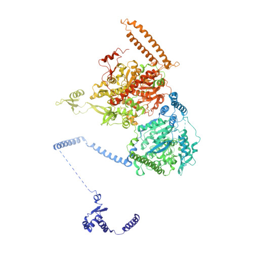
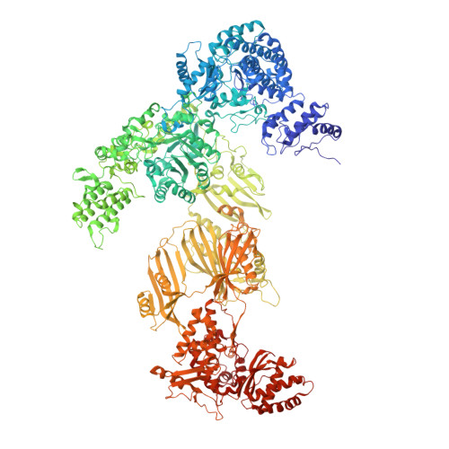
Electron cryomicroscopy observation of acyl carrier protein translocation in type I fungal fatty acid synthase.

Lou, J.W.Iyer, K.R.Hasan, S.M.N.Cowen, L.E.Mazhab-Jafari, M.T.

(2019) Sci Rep 9: 12987-12987

  • DOI: https://doi.org/10.1038/s41598-019-49261-3
  • Primary Citation of Related Structures:  
    6U5T, 6U5U, 6U5V, 6U5W

  • PubMed Abstract: 

    During fatty acid biosynthesis, acyl carrier proteins (ACPs) from type I fungal fatty acid synthase (FAS) shuttle substrates and intermediates within a reaction chamber that hosts multiple spatially-fixed catalytic centers. A major challenge in understanding the mechanism of ACP-mediated substrate shuttling is experimental observation of its transient interaction landscape within the reaction chamber. Here, we have shown that ACP spatial distribution is sensitive to the presence of substrates in a catalytically inhibited state, which enables high-resolution investigation of the ACP-dependent conformational transitions within the enoyl reductase (ER) reaction site. In two fungal FASs with distinct ACP localization, the shuttling domain is targeted to the ketoacyl-synthase (KS) domain and away from other catalytic centers, such as acetyl-transferase (AT) and ER domains by steric blockage of the KS active site followed by addition of substrates. These studies strongly suggest that acylation of phosphopantetheine arm of ACP may be an integral part of the substrate shuttling mechanism in type I fungal FAS.


  • Organizational Affiliation

    Department of Medical Biophysics, University of Toronto, Princess Margaret Cancer Research Institute, Toronto, Ontario, Canada.


Macromolecules
Find similar proteins by:  (by identity cutoff)  |  3D Structure
Entity ID: 1
MoleculeChains Sequence LengthOrganismDetailsImage
Fatty acid synthase subunit alpha1,722Candida albicansMutation(s): 0 
EC: 2.3.1.86 (PDB Primary Data), 1.1.1.100 (PDB Primary Data), 2.3.1.41 (PDB Primary Data)
UniProt
Find proteins for P43098 (Candida albicans)
Explore P43098 
Go to UniProtKB:  P43098
Entity Groups  
Sequence Clusters30% Identity50% Identity70% Identity90% Identity95% Identity100% Identity
UniProt GroupP43098
Sequence Annotations
Expand
  • Reference Sequence
Find similar proteins by:  (by identity cutoff)  |  3D Structure
Entity ID: 2
MoleculeChains Sequence LengthOrganismDetailsImage
Fatty acid synthase subunit beta2,037Candida albicansMutation(s): 0 
EC: 2.3.1.86 (PDB Primary Data), 4.2.1.59 (PDB Primary Data), 1.3.1.9 (PDB Primary Data), 2.3.1.38 (PDB Primary Data), 2.3.1.39 (PDB Primary Data), 3.1.2.14 (PDB Primary Data)
UniProt
Find proteins for P34731 (Candida albicans)
Explore P34731 
Go to UniProtKB:  P34731
Entity Groups  
Sequence Clusters30% Identity50% Identity70% Identity90% Identity95% Identity100% Identity
UniProt GroupP34731
Sequence Annotations
Expand
  • Reference Sequence
Experimental Data & Validation

Experimental Data

  • Method: ELECTRON MICROSCOPY
  • Resolution: 3.30 Å
  • Aggregation State: PARTICLE 
  • Reconstruction Method: SINGLE PARTICLE 
EM Software:
TaskSoftware PackageVersion
MODEL REFINEMENTPHENIX
RECONSTRUCTIONcryoSPARC2

Structure Validation

View Full Validation Report


Entry History 

Deposition Data

Revision History  (Full details and data files)

  • Version 1.0: 2019-10-16
    Type: Initial release
  • Version 1.1: 2024-03-20
    Changes: Data collection, Database references, Derived calculations, Refinement description