6U0O

Crystal structure of a peptidoglycan release complex, SagB-SpdC, in lipidic cubic phase


Experimental Data Snapshot

  • Method: X-RAY DIFFRACTION
  • Resolution: 2.60 Å
  • R-Value Free: 0.279 
  • R-Value Work: 0.253 
  • R-Value Observed: 0.255 

wwPDB Validation   3D Report Full Report


Ligand Structure Quality Assessment 


This is version 1.3 of the entry. See complete history


Literature

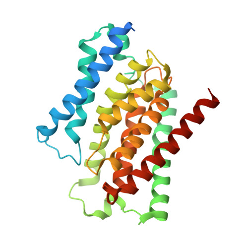
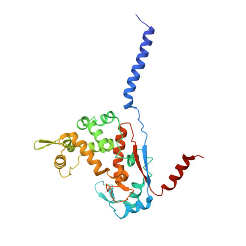
Structure and reconstitution of a hydrolase complex that may release peptidoglycan from the membrane after polymerization.

Schaefer, K.Owens, T.W.Page, J.E.Santiago, M.Kahne, D.Walker, S.

(2021) Nat Microbiol 6: 34-43

  • DOI: https://doi.org/10.1038/s41564-020-00808-5
  • Primary Citation of Related Structures:  
    6U0O

  • PubMed Abstract: 

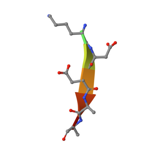
    Bacteria are encapsulated by a peptidoglycan cell wall that is essential for their survival 1 . During cell wall assembly, a lipid-linked disaccharide-peptide precursor called lipid II is polymerized and cross-linked to produce mature peptidoglycan. As lipid II is polymerized, nascent polymers remain membrane-anchored at one end, and the other end becomes cross-linked to the matrix 2-4 . How bacteria release newly synthesized peptidoglycan strands from the membrane to complete the synthesis of mature peptidoglycan is a long-standing question. Here, we show that a Staphylococcus aureus cell wall hydrolase and a membrane protein that contains eight transmembrane helices form a complex that may function as a peptidoglycan release factor. The complex cleaves nascent peptidoglycan internally to produce free oligomers as well as lipid-linked oligomers that can undergo further elongation. The polytopic membrane protein, which is similar to a eukaryotic CAAX protease, controls the length of these products. A structure of the complex at a resolution of 2.6 Å shows that the membrane protein scaffolds the hydrolase to orient its active site for cleaving the glycan strand. We propose that this complex functions to detach newly synthesized peptidoglycan polymer from the cell membrane to complete integration into the cell wall matrix.


  • Organizational Affiliation

    Department of Microbiology, Harvard Medical School, Boston, MA, USA.


Macromolecules
Find similar proteins by:  (by identity cutoff)  |  3D Structure
Entity ID: 1
MoleculeChains Sequence LengthOrganismDetailsImage
Lysostaphin resistance protein A278Staphylococcus aureus RF122Mutation(s): 0 
Gene Names: lyrASAB2212
Membrane Entity: Yes 
UniProt
Find proteins for Q2YYX7 (Staphylococcus aureus (strain bovine RF122 / ET3-1))
Explore Q2YYX7 
Go to UniProtKB:  Q2YYX7
Entity Groups  
Sequence Clusters30% Identity50% Identity70% Identity90% Identity95% Identity100% Identity
UniProt GroupQ2YYX7
Sequence Annotations
Expand
  • Reference Sequence
Find similar proteins by:  (by identity cutoff)  |  3D Structure
Entity ID: 2
MoleculeChains Sequence LengthOrganismDetailsImage
LYZ2 domain-containing protein297Staphylococcus aureus subsp. aureus NCTC 8325Mutation(s): 0 
Gene Names: SAOUHSC_01895
Membrane Entity: Yes 
UniProt
Find proteins for Q2FXF4 (Staphylococcus aureus (strain NCTC 8325 / PS 47))
Explore Q2FXF4 
Go to UniProtKB:  Q2FXF4
Entity Groups  
Sequence Clusters30% Identity50% Identity70% Identity90% Identity95% Identity100% Identity
UniProt GroupQ2FXF4
Sequence Annotations
Expand
  • Reference Sequence

Find similar proteins by:  Sequence   |   3D Structure  

Entity ID: 3
MoleculeChains Sequence LengthOrganismDetailsImage
FLAG peptideC [auth D]8synthetic constructMutation(s): 0 
Sequence Annotations
Expand
  • Reference Sequence
Small Molecules
Ligands 5 Unique
IDChains Name / Formula / InChI Key2D Diagram3D Interactions
OLC
Query on OLC

Download Ideal Coordinates CCD File 
F [auth A](2R)-2,3-dihydroxypropyl (9Z)-octadec-9-enoate
C21 H40 O4
RZRNAYUHWVFMIP-GDCKJWNLSA-N
PG4
Query on PG4

Download Ideal Coordinates CCD File 
E [auth A]TETRAETHYLENE GLYCOL
C8 H18 O5
UWHCKJMYHZGTIT-UHFFFAOYSA-N
FLC
Query on FLC

Download Ideal Coordinates CCD File 
D [auth A]CITRATE ANION
C6 H5 O7
KRKNYBCHXYNGOX-UHFFFAOYSA-K
AE3
Query on AE3

Download Ideal Coordinates CCD File 
J [auth B],
K [auth B]
2-(2-ETHOXYETHOXY)ETHANOL
C6 H14 O3
XXJWXESWEXIICW-UHFFFAOYSA-N
SO4
Query on SO4

Download Ideal Coordinates CCD File 
G [auth A],
H [auth B],
I [auth B]
SULFATE ION
O4 S
QAOWNCQODCNURD-UHFFFAOYSA-L
Experimental Data & Validation

Experimental Data

  • Method: X-RAY DIFFRACTION
  • Resolution: 2.60 Å
  • R-Value Free: 0.279 
  • R-Value Work: 0.253 
  • R-Value Observed: 0.255 
  • Space Group: C 1 2 1
Unit Cell:
Length ( Å )Angle ( ˚ )
a = 156.669α = 90
b = 54.758β = 107.18
c = 96.418γ = 90
Software Package:
Software NamePurpose
PHENIXrefinement
XDSdata reduction
Aimlessdata scaling
PHASERphasing
Cootmodel building

Structure Validation

View Full Validation Report


Ligand Structure Quality Assessment 


Entry History & Funding Information

Deposition Data


Funding OrganizationLocationGrant Number
National Institutes of Health/National Institute of General Medical Sciences (NIH/NIGMS)United States5R01GM076710-12

Revision History  (Full details and data files)

  • Version 1.0: 2020-09-23
    Type: Initial release
  • Version 1.1: 2020-11-25
    Changes: Database references
  • Version 1.2: 2021-01-06
    Changes: Database references
  • Version 1.3: 2023-10-11
    Changes: Data collection, Database references, Refinement description