6TYD

Structure of human LDB1 in complex with SSBP2


Experimental Data Snapshot

  • Method: X-RAY DIFFRACTION
  • Resolution: 2.80 Å
  • R-Value Free: 0.281 
  • R-Value Work: 0.240 
  • R-Value Observed: 0.244 

wwPDB Validation   3D Report Full Report


This is version 1.2 of the entry. See complete history


Literature

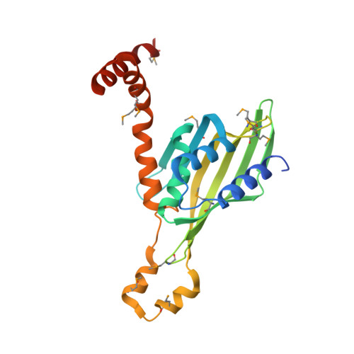
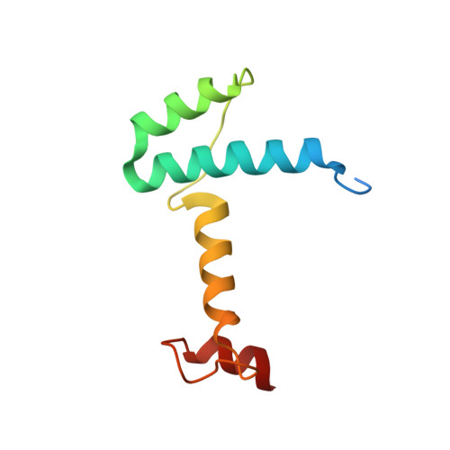
Crystal structure of human LDB1 in complex with SSBP2.

Wang, H.Kim, J.Wang, Z.Yan, X.X.Dean, A.Xu, W.

(2020) Proc Natl Acad Sci U S A 117: 1042-1048

  • DOI: https://doi.org/10.1073/pnas.1914181117
  • Primary Citation of Related Structures:  
    6TYD

  • PubMed Abstract: 

    The Lim domain binding proteins (LDB1 and LDB2 in human and Chip in Drosophila ) play critical roles in cell fate decisions through partnership with multiple Lim-homeobox and Lim-only proteins in diverse developmental systems including cardiogenesis, neurogenesis, and hematopoiesis. In mammalian erythroid cells, LDB1 dimerization supports long-range connections between enhancers and genes involved in erythropoiesis, including the β-globin genes. Single-stranded DNA binding proteins (SSBPs) interact specifically with the LDB/Chip conserved domain (LCCD) of LDB proteins and stabilize LDBs by preventing their proteasomal degradation, thus promoting their functions in gene regulation. The structural basis for LDB1 self-interaction and interface with SSBPs is unclear. Here we report a crystal structure of the human LDB1/SSBP2 complex at 2.8-Å resolution. The LDB1 dimerization domain (DD) contains an N-terminal nuclear transport factor 2 (NTF2)-like subdomain and a small helix 4-helix 5 subdomain, which together form the LDB1 dimerization interface. The 2 LCCDs in the symmetric LDB1 dimer flank the core DDs, with each LCCD forming extensive interactions with an SSBP2 dimer. The conserved linker between LDB1 DD and LCCD covers a potential ligand-binding pocket of the LDB1 NTF2-like subdomain and may serve as a regulatory site for LDB1 structure and function. Our structural and biochemical data provide a much-anticipated structural basis for understanding how LDB1 and the LDB1/SSBP interactions form the structural core of diverse complexes mediating cell choice decisions and long-range enhancer-promoter interactions.


  • Organizational Affiliation

    National Laboratory of Biomacromolecules, Chinese Academy of Sciences Center for Excellence in Biomacromolecules, Institute of Biophysics, Chinese Academy of Sciences, 100101 Beijing, China.


Macromolecules
Find similar proteins by:  (by identity cutoff)  |  3D Structure
Entity ID: 1
MoleculeChains Sequence LengthOrganismDetailsImage
LIM domain-binding protein 1A [auth V]231Homo sapiensMutation(s): 0 
Gene Names: LDB1CLIM2
UniProt & NIH Common Fund Data Resources
Find proteins for Q86U70 (Homo sapiens)
Explore Q86U70 
Go to UniProtKB:  Q86U70
PHAROS:  Q86U70
GTEx:  ENSG00000198728 
Entity Groups  
Sequence Clusters30% Identity50% Identity70% Identity90% Identity95% Identity100% Identity
UniProt GroupQ86U70
Sequence Annotations
Expand
  • Reference Sequence
Find similar proteins by:  (by identity cutoff)  |  3D Structure
Entity ID: 2
MoleculeChains Sequence LengthOrganismDetailsImage
Single-stranded DNA-binding protein 2B [auth A],
C [auth B]
95Homo sapiensMutation(s): 0 
Gene Names: SSBP2SSDP2
UniProt & NIH Common Fund Data Resources
Find proteins for P81877 (Homo sapiens)
Explore P81877 
Go to UniProtKB:  P81877
PHAROS:  P81877
GTEx:  ENSG00000145687 
Entity Groups  
Sequence Clusters30% Identity50% Identity70% Identity90% Identity95% Identity100% Identity
UniProt GroupP81877
Sequence Annotations
Expand
  • Reference Sequence
Small Molecules
Modified Residues  1 Unique
IDChains TypeFormula2D DiagramParent
MSE
Query on MSE
A [auth V]L-PEPTIDE LINKINGC5 H11 N O2 SeMET
Experimental Data & Validation

Experimental Data

  • Method: X-RAY DIFFRACTION
  • Resolution: 2.80 Å
  • R-Value Free: 0.281 
  • R-Value Work: 0.240 
  • R-Value Observed: 0.244 
  • Space Group: P 65 2 2
Unit Cell:
Length ( Å )Angle ( ˚ )
a = 104.322α = 90
b = 104.322β = 90
c = 250.419γ = 120
Software Package:
Software NamePurpose
PHENIXrefinement
PDB_EXTRACTdata extraction
HKL-2000data reduction
HKL-2000data scaling
PHENIXphasing

Structure Validation

View Full Validation Report


Entry History 

Deposition Data

Revision History  (Full details and data files)

  • Version 1.0: 2020-01-01
    Type: Initial release
  • Version 1.1: 2020-01-15
    Changes: Database references
  • Version 1.2: 2020-01-29
    Changes: Database references