6TFQ

Structure in P3212 form of the PBP/SBP MoaA in complex with mannopinic acid from A.tumefacien R10


Experimental Data Snapshot

  • Method: X-RAY DIFFRACTION
  • Resolution: 2.05 Å
  • R-Value Free: 0.199 
  • R-Value Work: 0.185 
  • R-Value Observed: 0.186 

wwPDB Validation   3D Report Full Report


Ligand Structure Quality Assessment 


This is version 1.2 of the entry. See complete history


Literature

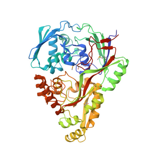
Import pathways of the mannityl-opines into the bacterial pathogen Agrobacterium tumefaciens: structural, affinity and in vivo approaches.

Vigouroux, A.Dore, J.Marty, L.Aumont-Nicaise, M.Legrand, P.Dessaux, Y.Vial, L.Morera, S.

(2020) Biochem J 477: 615-628

  • DOI: https://doi.org/10.1042/BCJ20190886
  • Primary Citation of Related Structures:  
    6TFQ, 6TFS, 6TFX, 6TG2, 6TG3

  • PubMed Abstract: 

    Agrobacterium tumefaciens pathogens use specific compounds denoted opines as nutrients in their plant tumor niche. These opines are produced by the host plant cells genetically modified by agrobacteria. They are imported into bacteria via solute-binding proteins (SBPs) in association with ATP-binding cassette transporters. The mannityl-opine family encompasses mannopine, mannopinic acid, agropine and agropinic acid. Structural and affinity data on mannopinic acid bound to SBPs are currently lacking while those of the three others mannityl opines are available. We investigated the molecular basis of two pathways for mannopinic acid uptake. MoaA was proposed as the specific SBP for mannopinic acid import in mannityl opines-assimilating agrobacteria, which was validated here using genetic studies and affinity measurements. We structurally characterized the mannopinic acid-binding mode of MoaA in two crystal forms at 2.05 and 1.57 Å resolution. We demonstrated that the non-specific SBP MotA, so far characterized as mannopine and Amadori compound importer, was also able to transport mannopinic acid. The structure of MotA bound to mannopinic acid at 2.2 Å resolution defines a different mannopinic acid-binding signature, similar to that of mannopine. Combining in vitro and in vivo approaches, this work allowed us to complete the characterization of the mannityl-opines assimilation pathways, highlighting the important role of two dual imports of agropinic and mannopinic acids. Our data shed new light on how the mannityl-opines contribute to the establishment of the ecological niche of agrobacteria from the early to the late stages of tumor development.


  • Organizational Affiliation

    Université Paris-Saclay, CEA, CNRS, Institute for Integrative Biology of the Cell (I2BC), 91198 Gif-sur-Yvette, France.


Macromolecules
Find similar proteins by:  (by identity cutoff)  |  3D Structure
Entity ID: 1
MoleculeChains Sequence LengthOrganismDetailsImage
ABC transporter substrate-binding protein
A, B
500Agrobacterium tumefaciensMutation(s): 0 
Gene Names: moaAddpAA6U90_18755At1D1609_52430AtA6_55190
UniProt
Find proteins for O50271 (Rhizobium radiobacter)
Explore O50271 
Go to UniProtKB:  O50271
Entity Groups  
Sequence Clusters30% Identity50% Identity70% Identity90% Identity95% Identity100% Identity
UniProt GroupO50271
Sequence Annotations
Expand
  • Reference Sequence
Small Molecules
Ligands 4 Unique
IDChains Name / Formula / InChI Key2D Diagram3D Interactions
N72 (Subject of Investigation/LOI)
Query on N72

Download Ideal Coordinates CCD File 
D [auth A],
U [auth B]
(2~{R})-2-[[(3~{R},4~{R},5~{S})-3,4,5,6-tetrakis(oxidanyl)-2-oxidanylidene-hexyl]amino]pentanedioic acid
C11 H19 N O9
PUFNBARRTADWAC-XWPZMVOTSA-N
GLC
Query on GLC

Download Ideal Coordinates CCD File 
C [auth A],
T [auth B]
alpha-D-glucopyranose
C6 H12 O6
WQZGKKKJIJFFOK-DVKNGEFBSA-N
SO4
Query on SO4

Download Ideal Coordinates CCD File 
AA [auth B]
BA [auth B]
CA [auth B]
DA [auth B]
E [auth A]
AA [auth B],
BA [auth B],
CA [auth B],
DA [auth B],
E [auth A],
EA [auth B],
F [auth A],
FA [auth B],
G [auth A],
GA [auth B],
H [auth A],
I [auth A],
J [auth A],
K [auth A],
L [auth A],
M [auth A],
N [auth A],
O [auth A],
P [auth A],
Q [auth A],
R [auth A],
V [auth B],
W [auth B],
X [auth B],
Y [auth B],
Z [auth B]
SULFATE ION
O4 S
QAOWNCQODCNURD-UHFFFAOYSA-L
CL
Query on CL

Download Ideal Coordinates CCD File 
HA [auth B],
S [auth A]
CHLORIDE ION
Cl
VEXZGXHMUGYJMC-UHFFFAOYSA-M
Modified Residues  1 Unique
IDChains TypeFormula2D DiagramParent
MSE
Query on MSE
A, B
L-PEPTIDE LINKINGC5 H11 N O2 SeMET
Binding Affinity Annotations 
IDSourceBinding Affinity
N72 Binding MOAD:  6TFQ Kd: 2.1 (nM) from 1 assay(s)
Experimental Data & Validation

Experimental Data

  • Method: X-RAY DIFFRACTION
  • Resolution: 2.05 Å
  • R-Value Free: 0.199 
  • R-Value Work: 0.185 
  • R-Value Observed: 0.186 
  • Space Group: P 32 1 2
Unit Cell:
Length ( Å )Angle ( ˚ )
a = 155.98α = 90
b = 155.98β = 90
c = 181.75γ = 120
Software Package:
Software NamePurpose
BUSTERrefinement
PDB_EXTRACTdata extraction
XDSdata reduction
XSCALEdata scaling
SHELXCDphasing

Structure Validation

View Full Validation Report


Ligand Structure Quality Assessment 


Entry History 

Deposition Data

Revision History  (Full details and data files)

  • Version 1.0: 2020-01-22
    Type: Initial release
  • Version 1.1: 2020-02-19
    Changes: Data collection, Database references
  • Version 1.2: 2020-07-29
    Type: Remediation
    Reason: Carbohydrate remediation
    Changes: Data collection, Derived calculations, Structure summary