6TC9

Crystal structure of MutM from Neisseria meningitidis


Experimental Data Snapshot

  • Method: X-RAY DIFFRACTION
  • Resolution: 2.17 Å
  • R-Value Free: 0.281 
  • R-Value Work: 0.238 
  • R-Value Observed: 0.240 

wwPDB Validation   3D Report Full Report


This is version 1.2 of the entry. See complete history


Literature

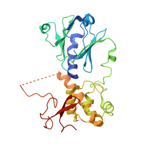
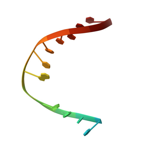
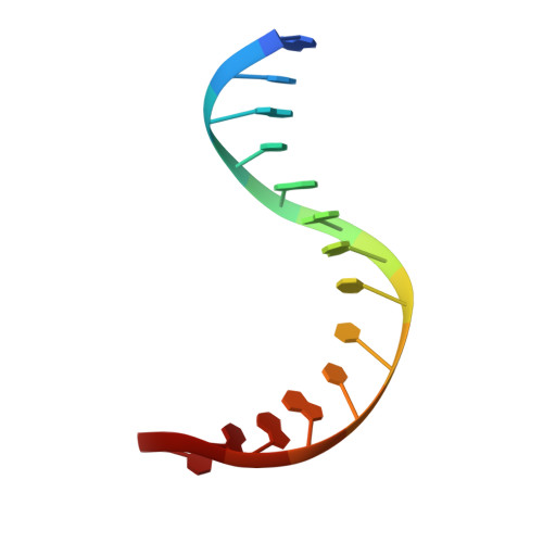
Conformational changes of DNA repair glycosylase MutM triggered by DNA binding.

Landova, B.Silhan, J.

(2020) FEBS Lett 594: 3032-3044

  • DOI: https://doi.org/10.1002/1873-3468.13876
  • Primary Citation of Related Structures:  
    6TC6, 6TC9

  • PubMed Abstract: 

    Bacterial MutM is a DNA repair glycosylase removing DNA damage generated from oxidative stress and, therefore, preventing mutations and genomic instability. MutM belongs to the Fpg/Nei family of prokaryotic enzymes sharing structural and functional similarities with their eukaryotic counterparts, for example, NEIL1-NEIL3. Here, we present two crystal structures of MutM from pathogenic Neisseria meningitidis: a MutM holoenzyme and MutM bound to DNA. The free enzyme exists in an open conformation, while upon binding to DNA, both the enzyme and DNA undergo substantial structural changes and domain rearrangement. Our data show that not only NEI glycosylases but also the MutMs undergo dramatic conformational changes. Moreover, crystallographic data support the previously published observations that MutM enzymes are rather flexible and dynamic molecules.


  • Organizational Affiliation

    Institute of Organic Chemistry and Biochemistry, Czech Academy of Sciences, Prague, Czech Republic.


Macromolecules

Find similar proteins by:  (by identity cutoff)  |  3D Structure
Entity ID: 2
MoleculeChains Sequence LengthOrganismDetailsImage
Formamidopyrimidine-DNA glycosylaseC [auth A],
E [auth C]
275Neisseria meningitidis alpha522Mutation(s): 0 
Gene Names: mutMfpgNMALPHA522_0971
EC: 3.2.2.23 (PDB Primary Data), 4.2.99.18 (PDB Primary Data)
UniProt
Find proteins for I4E596 (Neisseria meningitidis alpha522)
Explore I4E596 
Go to UniProtKB:  I4E596
Entity Groups  
Sequence Clusters30% Identity50% Identity70% Identity90% Identity95% Identity100% Identity
UniProt GroupI4E596
Sequence Annotations
Expand
  • Reference Sequence

Find similar nucleic acids by:  Sequence   |   3D Structure  

Entity ID: 1
MoleculeChains LengthOrganismImage
DNA containing abasic site analogueA [auth D],
B [auth G]
14synthetic construct
Sequence Annotations
Expand
  • Reference Sequence

Find similar nucleic acids by:  Sequence   |   3D Structure  

Entity ID: 3
MoleculeChains LengthOrganismImage
DNAD [auth B],
F
14synthetic construct
Sequence Annotations
Expand
  • Reference Sequence
Experimental Data & Validation

Experimental Data

  • Method: X-RAY DIFFRACTION
  • Resolution: 2.17 Å
  • R-Value Free: 0.281 
  • R-Value Work: 0.238 
  • R-Value Observed: 0.240 
  • Space Group: C 1 2 1
Unit Cell:
Length ( Å )Angle ( ˚ )
a = 130.77α = 90
b = 80.38β = 89.95
c = 83.62γ = 90
Software Package:
Software NamePurpose
PHENIXrefinement
XSCALEdata processing
PHASERphasing
XDSdata scaling
XDSdata reduction

Structure Validation

View Full Validation Report


Entry History & Funding Information

Deposition Data


Funding OrganizationLocationGrant Number
Czech Academy of SciencesCzech Republic17-21649Y

Revision History  (Full details and data files)

  • Version 1.0: 2020-09-16
    Type: Initial release
  • Version 1.1: 2020-12-16
    Changes: Database references
  • Version 1.2: 2024-01-24
    Changes: Data collection, Database references, Refinement description