Structure and molecular model refinement of Aspergillus oryzae (TAKA) alpha-amylase: an application of the simulated-annealing method.
Swift, H.J., Brady, L., Derewenda, Z.S., Dodson, E.J., Dodson, G.G., Turkenburg, J.P., Wilkinson, A.J.(1991) Acta Crystallogr B 47: 535-544
- PubMed: 1930835
- DOI: https://doi.org/10.1107/s0108768191001970
- Primary Citation of Related Structures:
6TAA - PubMed Abstract:
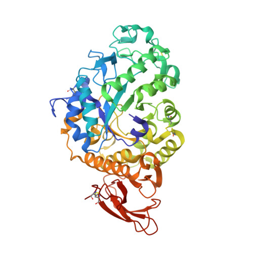
Monoclinic crystals of a neutral alpha-amylase from Aspergillus oryzae, containing three molecules in the asymmetric unit, have been reported previously and studied at 3 A resolution [Matsuura, Kunusoki, Harada & Kakudo (1984). J. Biochem. 95, 697-702]. Here we report the solution of the structure of this enzyme in a different crystal form (space group P2(1)2(1)2(1), a = 50.9, b = 67.2, c = 132.7 A), with only one molecule in the asymmetric unit. The structure was solved by the molecular replacement method, using a model of acid alpha-amylase from a related fungus A. niger [Brady, Brzozowski, Derewenda, Dodson & Dodson (1991). Acta Cryst. B47, 527-535]. Conventional least-squares crystallographic refinement failed to converge in a satisfactory manner, and the technique of molecular dynamics in the form of the XPLOR package [Brunger (1988). XPLOR Manual. Yale Univ., USA] was used to overcome the problem. A large rigid-body type movement of the C-terminal domain was identified and accounted for. The final round of restrained least-squares refinement (at 2.1 A resolution) including 3675 protein atoms and 247 water molecules resulted in a conventional crystallographic R factor of 0.183 and an atomic model which conforms well to standard stereochemical parameters (standard deviation of bond lengths from their expected values is 0.028 A, while that for planar groups is 0.029 A).
Organizational Affiliation:
Department of Chemistry, University of York, Heslington, England.