Structures of three MORN repeat proteins and a re-evaluation of the proposed lipid-binding properties of MORN repeats.
Sajko, S., Grishkovskaya, I., Kostan, J., Graewert, M., Setiawan, K., Trubestein, L., Niedermuller, K., Gehin, C., Sponga, A., Puchinger, M., Gavin, A.C., Leonard, T.A., Svergun, D.I., Smith, T.K., Morriswood, B., Djinovic-Carugo, K.(2020) PLoS One 15: e0242677-e0242677
- PubMed: 33296386
- DOI: https://doi.org/10.1371/journal.pone.0242677
- Primary Citation of Related Structures:
6T4D, 6T4R, 6T68, 6T69, 6T6Q - PubMed Abstract:
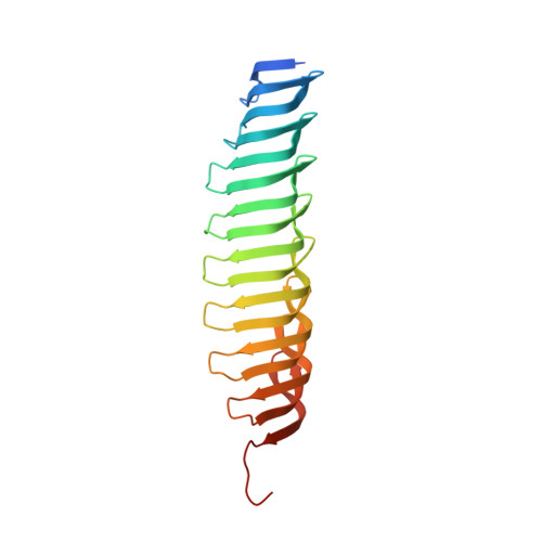
MORN (Membrane Occupation and Recognition Nexus) repeat proteins have a wide taxonomic distribution, being found in both prokaryotes and eukaryotes. Despite this ubiquity, they remain poorly characterised at both a structural and a functional level compared to other common repeats. In functional terms, they are often assumed to be lipid-binding modules that mediate membrane targeting. We addressed this putative activity by focusing on a protein composed solely of MORN repeats-Trypanosoma brucei MORN1. Surprisingly, no evidence for binding to membranes or lipid vesicles by TbMORN1 could be obtained either in vivo or in vitro. Conversely, TbMORN1 did interact with individual phospholipids. High- and low-resolution structures of the MORN1 protein from Trypanosoma brucei and homologous proteins from the parasites Toxoplasma gondii and Plasmodium falciparum were obtained using a combination of macromolecular crystallography, small-angle X-ray scattering, and electron microscopy. This enabled a first structure-based definition of the MORN repeat itself. Furthermore, all three structures dimerised via their C-termini in an antiparallel configuration. The dimers could form extended or V-shaped quaternary structures depending on the presence of specific interface residues. This work provides a new perspective on MORN repeats, showing that they are protein-protein interaction modules capable of mediating both dimerisation and oligomerisation.
Organizational Affiliation:
Department of Structural and Computational Biology, Max Perutz Labs, University of Vienna, Vienna, Austria.