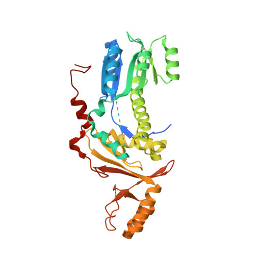
The structure of the Legionella response regulator LqsR reveals amino acids critical for phosphorylation and dimerization.
Hochstrasser, R., Hutter, C.A.J., Arnold, F.M., Barlocher, K., Seeger, M.A., Hilbi, H.(2020) Mol Microbiol 113: 1070-1084
- PubMed: 31997467
- DOI: https://doi.org/10.1111/mmi.14477
- Primary Citation of Related Structures:
6SY9 - PubMed Abstract:
The water-borne bacterium Legionella pneumophila replicates in environmental protozoa and upon inhalation destroys alveolar macrophages, thus causing a potentially fatal pneumonia termed 'Legionnaires' disease'. L. pneumophila employs the Legionella quorum sensing (Lqs) system to control its life cycle, pathogen-host cell interactions, motility and natural competence. Signaling through the Lqs system occurs through the α-hydroxyketone compound Legionella autoinducer-1 (LAI-1) and converges on the prototypic response regulator LqsR, which dimerizes upon phosphorylation of the conserved aspartate, D 108 . In this study, we determine the high-resolution structure of monomeric LqsR. The structure reveals a receiver domain adopting a canonical (βα) 5 fold, which is connected through an additional sixth helix and an extended α5-helix to a novel output domain. The two domains delineate a mainly positively charged groove, and the output domain adopts a five-stranded antiparallel β-sheet fold similar to nucleotide-binding proteins. Structure-based mutagenesis identified amino acids critical for LqsR phosphorylation and dimerization. Upon phosphorylation, the LqsR D172A and LqsR D302N/E303Q mutant proteins dimerized even more readily than wild-type LqsR, and no evidence for semi-phosphorylated heterodimers was obtained. Taken together, the high-resolution structure of LqsR reveals functionally relevant amino acid residues implicated in signal transduction of the prototypic response regulator.
Organizational Affiliation:
Institute of Medical Microbiology, University of Zürich, Zürich, Switzerland.