6SQG

Crystal structure of viral rhodopsin OLPVRII


Experimental Data Snapshot

  • Method: X-RAY DIFFRACTION
  • Resolution: 1.90 Å
  • R-Value Free: 0.210 
  • R-Value Work: 0.171 
  • R-Value Observed: 0.173 

wwPDB Validation   3D Report Full Report


Ligand Structure Quality Assessment 


This is version 1.2 of the entry. See complete history


Literature

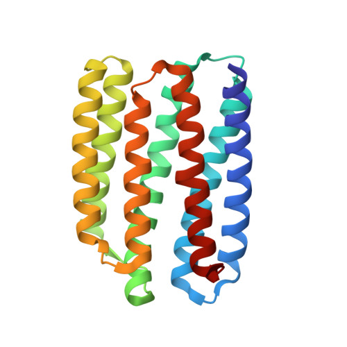
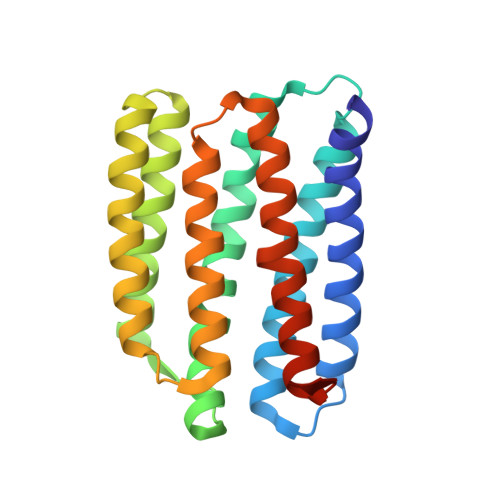
Unique structure and function of viral rhodopsins.

Bratanov, D.Kovalev, K.Machtens, J.P.Astashkin, R.Chizhov, I.Soloviov, D.Volkov, D.Polovinkin, V.Zabelskii, D.Mager, T.Gushchin, I.Rokitskaya, T.Antonenko, Y.Alekseev, A.Shevchenko, V.Yutin, N.Rosselli, R.Baeken, C.Borshchevskiy, V.Bourenkov, G.Popov, A.Balandin, T.Buldt, G.Manstein, D.J.Rodriguez-Valera, F.Fahlke, C.Bamberg, E.Koonin, E.Gordeliy, V.

(2019) Nat Commun 10: 4939-4939

  • DOI: https://doi.org/10.1038/s41467-019-12718-0
  • Primary Citation of Related Structures:  
    6SQG

  • PubMed Abstract: 

    Recently, two groups of rhodopsin genes were identified in large double-stranded DNA viruses. The structure and function of viral rhodopsins are unknown. We present functional characterization and high-resolution structure of an Organic Lake Phycodnavirus rhodopsin II (OLPVRII) of group 2. It forms a pentamer, with a symmetrical, bottle-like central channel with the narrow vestibule in the cytoplasmic part covered by a ring of 5 arginines, whereas 5 phenylalanines form a hydrophobic barrier in its exit. The proton donor E42 is placed in the helix B. The structure is unique among the known rhodopsins. Structural and functional data and molecular dynamics suggest that OLPVRII might be a light-gated pentameric ion channel analogous to pentameric ligand-gated ion channels, however, future patch clamp experiments should prove this directly. The data shed light on a fundamentally distinct branch of rhodopsins and may contribute to the understanding of virus-host interactions in ecologically important marine protists.


  • Organizational Affiliation

    Institute of Complex Systems (ICS), ICS-6: Structural Biochemistry, Forschungszentrum Jülich, Jülich, Germany.


Macromolecules
Find similar proteins by:  (by identity cutoff)  |  3D Structure
Entity ID: 1
MoleculeChains Sequence LengthOrganismDetailsImage
viral rhodopsin OLPVRII
A, B, C, D
211Organic Lake phycodnavirusMutation(s): 0 
Gene Names: 162286292
Membrane Entity: Yes 
UniProt
Find proteins for F2Y2Z0 (Organic Lake phycodnavirus)
Explore F2Y2Z0 
Go to UniProtKB:  F2Y2Z0
Entity Groups  
Sequence Clusters30% Identity50% Identity70% Identity90% Identity95% Identity100% Identity
UniProt GroupF2Y2Z0
Sequence Annotations
Expand
  • Reference Sequence
Find similar proteins by:  (by identity cutoff)  |  3D Structure
Entity ID: 2
MoleculeChains Sequence LengthOrganismDetailsImage
viral rhodopsin OLPVRII219Organic Lake phycodnavirusMutation(s): 0 
Gene Names: 162286292
Membrane Entity: Yes 
UniProt
Find proteins for F2Y2Z0 (Organic Lake phycodnavirus)
Explore F2Y2Z0 
Go to UniProtKB:  F2Y2Z0
Entity Groups  
Sequence Clusters30% Identity50% Identity70% Identity90% Identity95% Identity100% Identity
UniProt GroupF2Y2Z0
Sequence Annotations
Expand
  • Reference Sequence
Small Molecules
Ligands 3 Unique
IDChains Name / Formula / InChI Key2D Diagram3D Interactions
OLB
Query on OLB

Download Ideal Coordinates CCD File 
CB [auth D],
PB [auth E]
(2S)-2,3-dihydroxypropyl (9Z)-octadec-9-enoate
C21 H40 O4
RZRNAYUHWVFMIP-QJRAZLAKSA-N
RET
Query on RET

Download Ideal Coordinates CCD File 
BB [auth C],
EC [auth E],
MA [auth B],
OB [auth D],
W [auth A]
RETINAL
C20 H28 O
NCYCYZXNIZJOKI-OVSJKPMPSA-N
LFA
Query on LFA

Download Ideal Coordinates CCD File 
AA [auth B]
AB [auth C]
AC [auth E]
BA [auth B]
BC [auth E]
AA [auth B],
AB [auth C],
AC [auth E],
BA [auth B],
BC [auth E],
CA [auth B],
CC [auth E],
DA [auth B],
DB [auth D],
DC [auth E],
EA [auth B],
EB [auth D],
F [auth A],
FA [auth B],
FB [auth D],
G [auth A],
GA [auth B],
GB [auth D],
H [auth A],
HA [auth B],
HB [auth D],
I [auth A],
IA [auth B],
IB [auth D],
J [auth A],
JA [auth B],
JB [auth D],
K [auth A],
KA [auth B],
KB [auth D],
L [auth A],
LA [auth B],
LB [auth D],
M [auth A],
MB [auth D],
N [auth A],
NA [auth C],
NB [auth D],
O [auth A],
OA [auth C],
P [auth A],
PA [auth C],
Q [auth A],
QA [auth C],
QB [auth E],
R [auth A],
RA [auth C],
RB [auth E],
S [auth A],
SA [auth C],
SB [auth E],
T [auth A],
TA [auth C],
TB [auth E],
U [auth A],
UA [auth C],
UB [auth E],
V [auth A],
VA [auth C],
VB [auth E],
WA [auth C],
WB [auth E],
X [auth B],
XA [auth C],
XB [auth E],
Y [auth B],
YA [auth C],
YB [auth E],
Z [auth B],
ZA [auth C],
ZB [auth E]
EICOSANE
C20 H42
CBFCDTFDPHXCNY-UHFFFAOYSA-N
Experimental Data & Validation

Experimental Data

  • Method: X-RAY DIFFRACTION
  • Resolution: 1.90 Å
  • R-Value Free: 0.210 
  • R-Value Work: 0.171 
  • R-Value Observed: 0.173 
  • Space Group: P 1 21 1
Unit Cell:
Length ( Å )Angle ( ˚ )
a = 79.582α = 90
b = 99.699β = 117.17
c = 82.958γ = 90
Software Package:
Software NamePurpose
Aimlessdata scaling
REFMACrefinement
PDB_EXTRACTdata extraction
XDSdata reduction
MOLREPphasing

Structure Validation

View Full Validation Report


Ligand Structure Quality Assessment 


Entry History & Funding Information

Deposition Data


Funding OrganizationLocationGrant Number
French National Research AgencyFranceANR-15-CE11-0029-02/FA 301/11-1
French National Research AgencyFranceANR-10-INBS-05-02
Grenoble Alliance for Integrated Structural Cell BiologyFranceANR-17-EURE-0003
Russian Foundation for Basic ResearchRussian Federation19-52-15017
French National Research AgencyFranceANR-14-CE09-0028

Revision History  (Full details and data files)

  • Version 1.0: 2019-11-06
    Type: Initial release
  • Version 1.1: 2019-11-13
    Changes: Data collection, Database references
  • Version 1.2: 2024-01-24
    Changes: Data collection, Database references, Refinement description