Structural Characterization of EnpA D,L-Endopeptidase from Enterococcus faecalis Prophage Provides Insights into Substrate Specificity of M23 Peptidases.
Malecki, P.H., Mitkowski, P., Jagielska, E., Trochimiak, K., Mesnage, S., Sabala, I.(2021) Int J Mol Sci 22
- PubMed: 34281200
- DOI: https://doi.org/10.3390/ijms22137136
- Primary Citation of Related Structures:
6SMK - PubMed Abstract:
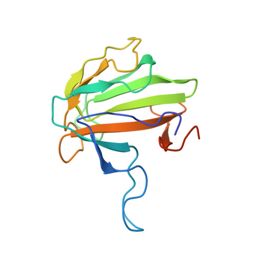
The best-characterized members of the M23 family are glycyl-glycine hydrolases, such as lysostaphin (Lss) from Staphylococcus simulans or LytM from Staphylococcus aureus . Recently, enzymes with broad specificities were reported, such as EnpA CD from Enterococcus faecalis , that cleaves D,L peptide bond between the stem peptide and a cross-bridge. Previously, the activity of EnpA CD was demonstrated only on isolated peptidoglycan fragments. Herein we report conditions in which EnpA CD lyses bacterial cells live with very high efficiency demonstrating great bacteriolytic potential, though limited to a low ionic strength environment. We have solved the structure of the EnpA CD H109A inactive variant and analyzed it in the context of related peptidoglycan hydrolases structures to reveal the bases for the specificity determination. All M23 structures share a very conserved β-sheet core which constitutes the rigid bottom of the substrate-binding groove and active site, while variable loops create the walls of the deep and narrow binding cleft. A detailed analysis of the binding groove architecture, specificity of M23 enzymes and D,L peptidases demonstrates that the substrate groove, which is particularly deep and narrow, is accessible preferably for peptides composed of amino acids with short side chains or subsequent L and D-isomers. As a result, the bottom of the groove is involved in interactions with the main chain of the substrate while the side chains are protruding in one plane towards the groove opening. We concluded that the selectivity of the substrates is based on their conformations allowed only for polyglycine chains and alternating chirality of the amino acids.
Organizational Affiliation:
International Institute of Molecular and Cell Biology, 02-109 Warsaw, Poland.