6SKI

The Tle hydrolase bound to the TTR domain of the VgrG spike of the Type 6 secretion system


Experimental Data Snapshot

  • Method: ELECTRON MICROSCOPY
  • Resolution: 2.60 Å
  • Aggregation State: PARTICLE 
  • Reconstruction Method: SINGLE PARTICLE 

wwPDB Validation   3D Report Full Report


This is version 1.1 of the entry. See complete history


Literature

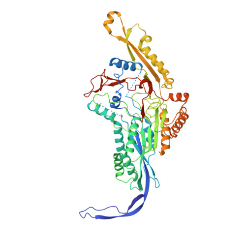
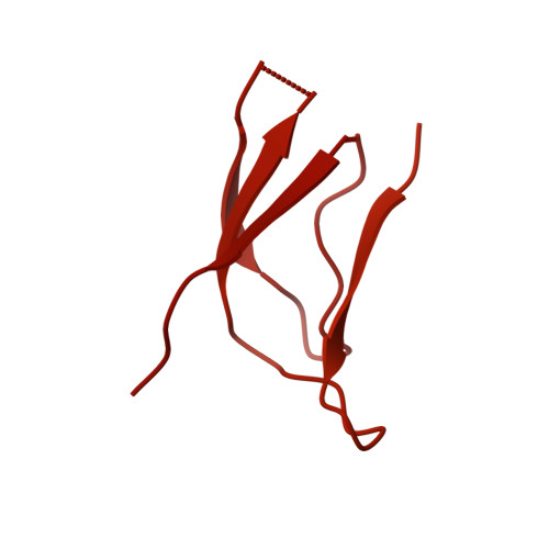
Structural basis for loading and inhibition of a bacterial T6SS phospholipase effector by the VgrG spike.

Flaugnatti, N.Rapisarda, C.Rey, M.Beauvois, S.G.Nguyen, V.A.Canaan, S.Durand, E.Chamot-Rooke, J.Cascales, E.Fronzes, R.Journet, L.

(2020) EMBO J 39: e104129-e104129

  • DOI: https://doi.org/10.15252/embj.2019104129
  • Primary Citation of Related Structures:  
    6SJL, 6SK0, 6SKI

  • PubMed Abstract: 

    The bacterial type VI secretion system (T6SS) is a macromolecular machine that injects effectors into prokaryotic and eukaryotic cells. The mode of action of the T6SS is similar to contractile phages: the contraction of a sheath structure pushes a tube topped by a spike into target cells. Effectors are loaded onto the spike or confined into the tube. In enteroaggregative Escherichia coli, the Tle1 phospholipase binds the C-terminal extension of the VgrG trimeric spike. Here, we purify the VgrG-Tle1 complex and show that a VgrG trimer binds three Tle1 monomers and inhibits their activity. Using covalent cross-linking coupled to high-resolution mass spectrometry, we provide information on the sites of contact and further identify the requirement for a Tle1 N-terminal secretion sequence in complex formation. Finally, we report the 2.6-Å-resolution cryo-electron microscopy tri-dimensional structure of the (VgrG) 3 -(Tle1) 3 complex revealing how the effector binds its cargo, and how VgrG inhibits Tle1 phospholipase activity. The inhibition of Tle1 phospholipase activity once bound to VgrG suggests that Tle1 dissociation from VgrG is required upon delivery.


  • Organizational Affiliation

    Laboratoire d'Ingénierie des Systèmes Macromoléculaires, Institut de Microbiologie de la Méditerranée, Aix-Marseille Université-CNRS UMR7255, Marseille, France.


Macromolecules
Find similar proteins by:  (by identity cutoff)  |  3D Structure
Entity ID: 1
MoleculeChains Sequence LengthOrganismDetailsImage
Putative type VI secretion proteinA [auth F]560Escherichia coliMutation(s): 0 
Gene Names: NCTC12950_00913NCTC9044_02520
UniProt
Find proteins for A0A377LA80 (Escherichia coli)
Explore A0A377LA80 
Go to UniProtKB:  A0A377LA80
Entity Groups  
Sequence Clusters30% Identity50% Identity70% Identity90% Identity95% Identity100% Identity
UniProt GroupA0A377LA80
Sequence Annotations
Expand
  • Reference Sequence
Find similar proteins by:  (by identity cutoff)  |  3D Structure
Entity ID: 2
MoleculeChains Sequence LengthOrganismDetailsImage
Putative type VI secretion proteinB [auth A]841Escherichia coliMutation(s): 0 
Gene Names: vgrGDL654_23170DM280_18435EF082_25505EHD42_22735EIT12_22235EIT27_22025NCTC9044_02519
UniProt
Find proteins for A0A3W2RZ19 (Escherichia coli)
Explore A0A3W2RZ19 
Go to UniProtKB:  A0A3W2RZ19
Entity Groups  
Sequence Clusters30% Identity50% Identity70% Identity90% Identity95% Identity100% Identity
UniProt GroupA0A3W2RZ19
Sequence Annotations
Expand
  • Reference Sequence
Experimental Data & Validation

Experimental Data

  • Method: ELECTRON MICROSCOPY
  • Resolution: 2.60 Å
  • Aggregation State: PARTICLE 
  • Reconstruction Method: SINGLE PARTICLE 
EM Software:
TaskSoftware PackageVersion
MODEL REFINEMENTPHENIX
RECONSTRUCTIONcryoSPARC

Structure Validation

View Full Validation Report


Entry History & Funding Information

Deposition Data


Funding OrganizationLocationGrant Number
French National Research AgencyFrance--
European Communitys Seventh Framework ProgrammeGermany653706

Revision History  (Full details and data files)

  • Version 1.0: 2020-04-15
    Type: Initial release
  • Version 1.1: 2020-10-28
    Changes: Database references