6SG8

Structure of Sosuga virus receptor binding protein

  • Classification: VIRAL PROTEIN
  • Organism(s): Sosuga virus
  • Expression System: Homo sapiens
  • Mutation(s): No 

  • Deposited: 2019-08-02 Released: 2019-10-09 
  • Deposition Author(s): Bowden, T.A., Stelfox, A.J.
  • Funding Organization(s): Medical Research Council (United Kingdom), Engineering and Physical Sciences Research Council, Wellcome Trust, National Institutes of Health/National Institute of General Medical Sciences (NIH/NIGMS)

Experimental Data Snapshot

  • Method: X-RAY DIFFRACTION
  • Resolution: 2.50 Å
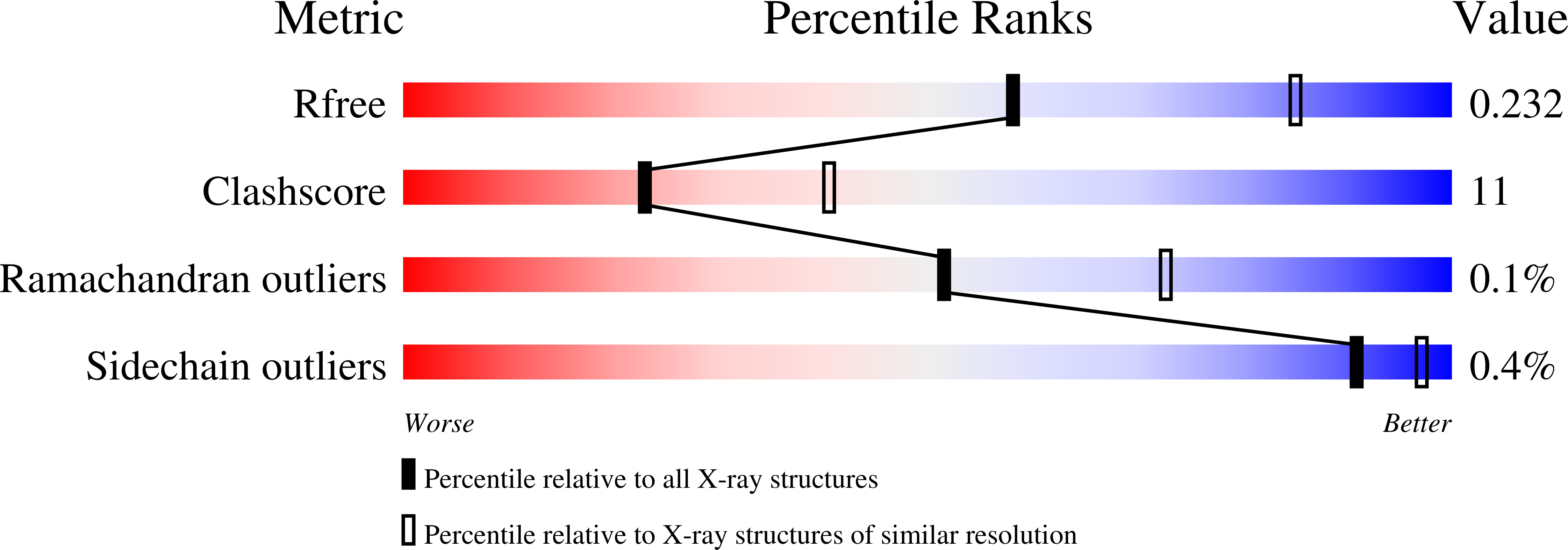
  • R-Value Free: 0.231 
  • R-Value Work: 0.194 
  • R-Value Observed: 0.196 

wwPDB Validation   3D Report Full Report


This is version 1.5 of the entry. See complete history


Literature

A structure-based rationale for sialic acid independent host-cell entry of Sosuga virus.

Stelfox, A.J.Bowden, T.A.

(2019) Proc Natl Acad Sci U S A 116: 21514-21520

  • DOI: https://doi.org/10.1073/pnas.1906717116
  • Primary Citation of Related Structures:  
    6SG8

  • PubMed Abstract: 

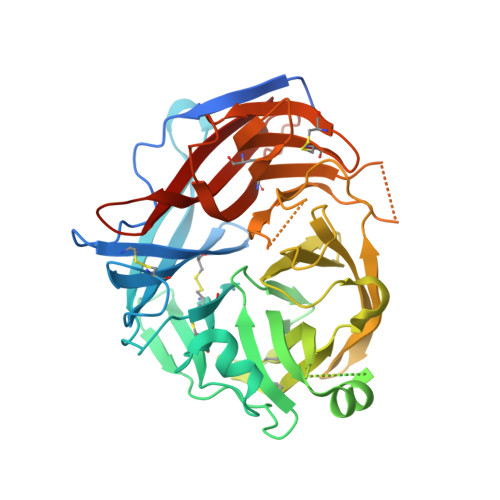
    The bat-borne paramyxovirus, Sosuga virus (SosV), is one of many paramyxoviruses recently identified and classified within the newly established genus Pararubulavirus , family Paramyxoviridae The envelope surface of SosV presents a receptor-binding protein (RBP), SosV-RBP, which facilitates host-cell attachment and entry. Unlike closely related hemagglutinin neuraminidase RBPs from other genera of the Paramyxoviridae , SosV-RBP and other pararubulavirus RBPs lack many of the stringently conserved residues required for sialic acid recognition and hydrolysis. We determined the crystal structure of the globular head region of SosV-RBP, revealing that while the glycoprotein presents a classical paramyxoviral six-bladed β-propeller fold and structurally classifies in close proximity to paramyxoviral RBPs with hemagglutinin-neuraminidase (HN) functionality, it presents a receptor-binding face incongruent with sialic acid recognition. Hemadsorption and neuraminidase activity analysis confirms the limited capacity of SosV-RBP to interact with sialic acid in vitro and indicates that SosV-RBP undergoes a nonclassical route of host-cell entry. The close overall structural conservation of SosV-RBP with other classical HN RBPs supports a model by which pararubulaviruses only recently diverged from sialic acid binding functionality.


  • Organizational Affiliation

    Division of Structural Biology, Wellcome Centre for Human Genetics, University of Oxford, OX3 7BN Oxford, United Kingdom.


Macromolecules
Find similar proteins by:  (by identity cutoff)  |  3D Structure
Entity ID: 1
MoleculeChains Sequence LengthOrganismDetailsImage
Hemagglutinin-neuraminidase
A, B
468Sosuga virusMutation(s): 0 
UniProt
Find proteins for W5SB61 (Sosuga virus)
Explore W5SB61 
Go to UniProtKB:  W5SB61
Entity Groups  
Sequence Clusters30% Identity50% Identity70% Identity90% Identity95% Identity100% Identity
UniProt GroupW5SB61
Sequence Annotations
Expand
  • Reference Sequence
Small Molecules
Ligands 3 Unique
IDChains Name / Formula / InChI Key2D Diagram3D Interactions
NAG
Query on NAG

Download Ideal Coordinates CCD File 
C [auth A]
D [auth A]
E [auth A]
F [auth A]
L [auth B]
C [auth A],
D [auth A],
E [auth A],
F [auth A],
L [auth B],
M [auth B],
N [auth B],
O [auth B],
P [auth B],
Q [auth B]
2-acetamido-2-deoxy-beta-D-glucopyranose
C8 H15 N O6
OVRNDRQMDRJTHS-FMDGEEDCSA-N
ACT
Query on ACT

Download Ideal Coordinates CCD File 
G [auth A]
H [auth A]
I [auth A]
J [auth A]
R [auth B]
G [auth A],
H [auth A],
I [auth A],
J [auth A],
R [auth B],
S [auth B],
T [auth B],
U [auth B],
V [auth B],
W [auth B]
ACETATE ION
C2 H3 O2
QTBSBXVTEAMEQO-UHFFFAOYSA-M
CA
Query on CA

Download Ideal Coordinates CCD File 
K [auth A],
X [auth B]
CALCIUM ION
Ca
BHPQYMZQTOCNFJ-UHFFFAOYSA-N
Experimental Data & Validation

Experimental Data

  • Method: X-RAY DIFFRACTION
  • Resolution: 2.50 Å
  • R-Value Free: 0.231 
  • R-Value Work: 0.194 
  • R-Value Observed: 0.196 
  • Space Group: P 1 21 1
Unit Cell:
Length ( Å )Angle ( ˚ )
a = 75.7α = 90
b = 84.04β = 112.85
c = 81.69γ = 90
Software Package:
Software NamePurpose
PHENIXrefinement
xia2data reduction
xia2data scaling
PHENIXphasing

Structure Validation

View Full Validation Report


Entry History & Funding Information

Deposition Data


Funding OrganizationLocationGrant Number
Medical Research Council (United Kingdom)United KingdomMR/L009528/1
Medical Research Council (United Kingdom)United KingdomMR/S007555/1
Engineering and Physical Sciences Research CouncilUnited KingdomEP/K503113/1
Engineering and Physical Sciences Research CouncilUnited KingdomEP/L505031/1
Engineering and Physical Sciences Research CouncilUnited KingdomEP/M50659X/1
Engineering and Physical Sciences Research CouncilUnited KingdomEP/M508111/1
Wellcome TrustUnited Kingdom203141/Z/16/Z
National Institutes of Health/National Institute of General Medical Sciences (NIH/NIGMS)United StatesAI123449

Revision History  (Full details and data files)

  • Version 1.0: 2019-10-09
    Type: Initial release
  • Version 1.1: 2019-10-23
    Changes: Data collection, Database references
  • Version 1.2: 2019-10-30
    Changes: Data collection, Database references
  • Version 1.3: 2020-07-29
    Type: Remediation
    Reason: Carbohydrate remediation
    Changes: Data collection, Derived calculations, Structure summary
  • Version 1.4: 2022-03-30
    Changes: Author supporting evidence, Database references, Structure summary
  • Version 1.5: 2024-01-24
    Changes: Data collection, Refinement description