6SDK

Crystal structure of bacterial ParB dimer bound to CDP


Experimental Data Snapshot

  • Method: X-RAY DIFFRACTION
  • Resolution: 1.81 Å
  • R-Value Free: 0.213 
  • R-Value Work: 0.184 
  • R-Value Observed: 0.185 

wwPDB Validation   3D Report Full Report


Ligand Structure Quality Assessment 


This is version 1.3 of the entry. See complete history


Literature

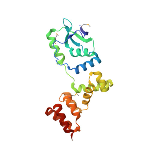
Self-organization ofparScentromeres by the ParB CTP hydrolase.

Soh, Y.M.Davidson, I.F.Zamuner, S.Basquin, J.Bock, F.P.Taschner, M.Veening, J.W.De Los Rios, P.Peters, J.M.Gruber, S.

(2019) Science 366: 1129-1133

  • DOI: https://doi.org/10.1126/science.aay3965
  • Primary Citation of Related Structures:  
    6SDK

  • PubMed Abstract: 

    ParABS systems facilitate chromosome segregation and plasmid partitioning in bacteria and archaea. ParB protein binds centromeric parS DNA sequences and spreads to flanking DNA. We show that ParB is an enzyme that hydrolyzes cytidine triphosphate (CTP) to cytidine diphosphate (CDP). parS DNA stimulates cooperative CTP binding by ParB and CTP hydrolysis. A nucleotide cocrystal structure elucidates the catalytic center of the dimerization-dependent ParB CTPase. Single-molecule imaging and biochemical assays recapitulate features of ParB spreading from parS in the presence but not absence of CTP. These findings suggest that centromeres assemble by self-loading of ParB DNA sliding clamps at parS ParB CTPase is not related to known nucleotide hydrolases and might be a promising target for developing new classes of antibiotics.


  • Organizational Affiliation

    Department of Fundamental Microbiology (DMF), Faculty of Biology and Medicine (FBM), University of Lausanne (UNIL), Lausanne, Switzerland.


Macromolecules
Find similar proteins by:  (by identity cutoff)  |  3D Structure
Entity ID: 1
MoleculeChains Sequence LengthOrganismDetailsImage
Stage 0 sporulation protein J
A, B, C, D
199Bacillus subtilis subsp. subtilis str. 168Mutation(s): 0 
Gene Names: spo0JBSU40960
UniProt
Find proteins for P26497 (Bacillus subtilis (strain 168))
Explore P26497 
Go to UniProtKB:  P26497
Entity Groups  
Sequence Clusters30% Identity50% Identity70% Identity90% Identity95% Identity100% Identity
UniProt GroupP26497
Sequence Annotations
Expand
  • Reference Sequence
Small Molecules
Ligands 2 Unique
IDChains Name / Formula / InChI Key2D Diagram3D Interactions
CDP
Query on CDP

Download Ideal Coordinates CCD File 
E [auth A],
H [auth B],
K [auth C],
N [auth D]
CYTIDINE-5'-DIPHOSPHATE
C9 H15 N3 O11 P2
ZWIADYZPOWUWEW-XVFCMESISA-N
CA
Query on CA

Download Ideal Coordinates CCD File 
F [auth A]
G [auth A]
I [auth B]
J [auth B]
L [auth C]
F [auth A],
G [auth A],
I [auth B],
J [auth B],
L [auth C],
M [auth C],
O [auth D],
P [auth D]
CALCIUM ION
Ca
BHPQYMZQTOCNFJ-UHFFFAOYSA-N
Modified Residues  1 Unique
IDChains TypeFormula2D DiagramParent
MSE
Query on MSE
A, B, C, D
L-PEPTIDE LINKINGC5 H11 N O2 SeMET
Experimental Data & Validation

Experimental Data

  • Method: X-RAY DIFFRACTION
  • Resolution: 1.81 Å
  • R-Value Free: 0.213 
  • R-Value Work: 0.184 
  • R-Value Observed: 0.185 
  • Space Group: P 1 21 1
Unit Cell:
Length ( Å )Angle ( ˚ )
a = 47.186α = 90
b = 104.501β = 102.568
c = 103.193γ = 90
Software Package:
Software NamePurpose
PHENIXrefinement
XDSdata reduction
XDSdata scaling
CRANK2phasing

Structure Validation

View Full Validation Report


Ligand Structure Quality Assessment 


Entry History & Funding Information

Deposition Data


Funding OrganizationLocationGrant Number
European Research CouncilSwitzerland724482

Revision History  (Full details and data files)

  • Version 1.0: 2019-10-23
    Type: Initial release
  • Version 1.1: 2019-10-30
    Changes: Data collection, Database references
  • Version 1.2: 2019-11-06
    Changes: Data collection, Database references
  • Version 1.3: 2019-12-11
    Changes: Database references