6S67

Structure of the Fluorescent Protein AausFP1 from Aequorea cf. australis at pH 7.0


Experimental Data Snapshot

  • Method: X-RAY DIFFRACTION
  • Resolution: 2.47 Å
  • R-Value Free: 0.228 
  • R-Value Work: 0.182 
  • R-Value Observed: 0.185 

wwPDB Validation   3D Report Full Report


This is version 1.3 of the entry. See complete history


Literature

Aequorea's secrets revealed: New fluorescent proteins with unique properties for bioimaging and biosensing.

Lambert, G.G.Depernet, H.Gotthard, G.Schultz, D.T.Navizet, I.Lambert, T.Adams, S.R.Torreblanca-Zanca, A.Chu, M.Bindels, D.S.Levesque, V.Nero Moffatt, J.Salih, A.Royant, A.Shaner, N.C.

(2020) PLoS Biol 18: e3000936-e3000936

  • DOI: https://doi.org/10.1371/journal.pbio.3000936
  • Primary Citation of Related Structures:  
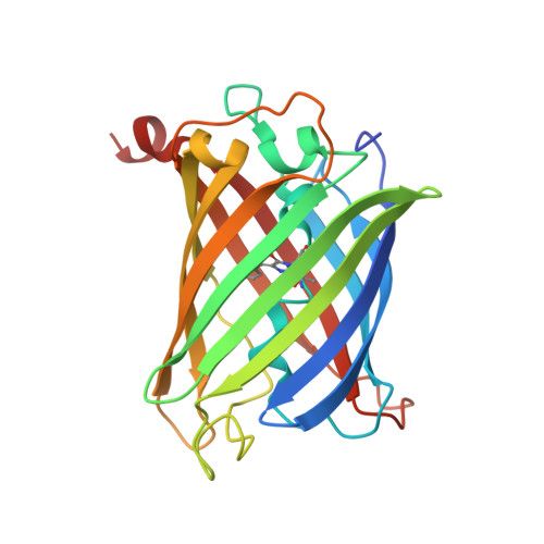
    6S67, 6S68

  • PubMed Abstract: 

    Using mRNA sequencing and de novo transcriptome assembly, we identified, cloned, and characterized 9 previously undiscovered fluorescent protein (FP) homologs from Aequorea victoria and a related Aequorea species, with most sequences highly divergent from A. victoria green fluorescent protein (avGFP). Among these FPs are the brightest green fluorescent protein (GFP) homolog yet characterized and a reversibly photochromic FP that responds to UV and blue light. Beyond green emitters, Aequorea species express purple- and blue-pigmented chromoproteins (CPs) with absorbances ranging from green to far-red, including 2 that are photoconvertible. X-ray crystallography revealed that Aequorea CPs contain a chemically novel chromophore with an unexpected crosslink to the main polypeptide chain. Because of the unique attributes of several of these newly discovered FPs, we expect that Aequorea will, once again, give rise to an entirely new generation of useful probes for bioimaging and biosensing.


  • Organizational Affiliation

    Department of Neurosciences, Center for Research in Biological Systems, University of California San Diego School of Medicine, La Jolla, California, United States of America.


Macromolecules
Find similar proteins by:  (by identity cutoff)  |  3D Structure
Entity ID: 1
MoleculeChains Sequence LengthOrganismDetailsImage
Aequorea cf. australis fluorescent protein 1 (AausFP1)
A, B, C, D
230Aequorea australisMutation(s): 0 
UniProt
Find proteins for A0A5J6CYI5 (Aequorea australis)
Explore A0A5J6CYI5 
Go to UniProtKB:  A0A5J6CYI5
Entity Groups  
Sequence Clusters30% Identity50% Identity70% Identity90% Identity95% Identity100% Identity
UniProt GroupA0A5J6CYI5
Sequence Annotations
Expand
  • Reference Sequence
Experimental Data & Validation

Experimental Data

  • Method: X-RAY DIFFRACTION
  • Resolution: 2.47 Å
  • R-Value Free: 0.228 
  • R-Value Work: 0.182 
  • R-Value Observed: 0.185 
  • Space Group: P 21 21 21
Unit Cell:
Length ( Å )Angle ( ˚ )
a = 65.08α = 90
b = 101.41β = 90
c = 161.43γ = 90
Software Package:
Software NamePurpose
REFMACrefinement
XDSdata reduction
XSCALEdata scaling
PHASERphasing
PDB_EXTRACTdata extraction

Structure Validation

View Full Validation Report


Entry History & Funding Information

Deposition Data


Funding OrganizationLocationGrant Number
National Institutes of Health/National Institute of General Medical Sciences (NIH/NIGMS)United States1R01GM109984-01

Revision History  (Full details and data files)

  • Version 1.0: 2020-07-22
    Type: Initial release
  • Version 1.1: 2021-02-03
    Changes: Database references
  • Version 1.2: 2022-03-30
    Changes: Author supporting evidence, Database references
  • Version 1.3: 2024-01-24
    Changes: Data collection, Refinement description