6S2V

Structure of the N-terminal catalytic region of T. thermophilus Rel


Experimental Data Snapshot

  • Method: X-RAY DIFFRACTION
  • Resolution: 2.96 Å
  • R-Value Free: 0.275 
  • R-Value Work: 0.221 
  • R-Value Observed: 0.224 

wwPDB Validation   3D Report Full Report


This is version 1.3 of the entry. See complete history


Literature

A nucleotide-switch mechanism mediates opposing catalytic activities of Rel enzymes.

Tamman, H.Van Nerom, K.Takada, H.Vandenberk, N.Scholl, D.Polikanov, Y.Hofkens, J.Talavera, A.Hauryliuk, V.Hendrix, J.Garcia-Pino, A.

(2020) Nat Chem Biol 16: 834-840

  • DOI: https://doi.org/10.1038/s41589-020-0520-2
  • Primary Citation of Related Structures:  
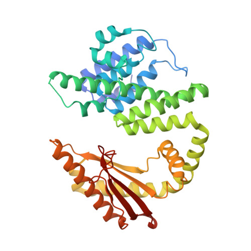
    6S2T, 6S2U, 6S2V

  • PubMed Abstract: 

    Bifunctional Rel stringent factors, the most abundant class of RelA/SpoT homologs, are ribosome-associated enzymes that transfer a pyrophosphate from ATP onto the 3' of guanosine tri-/diphosphate (GTP/GDP) to synthesize the bacterial alarmone (p)ppGpp, and also catalyze the 3' pyrophosphate hydrolysis to degrade it. The regulation of the opposing activities of Rel enzymes is a complex allosteric mechanism that remains an active research topic despite decades of research. We show that a guanine-nucleotide-switch mechanism controls catalysis by Thermus thermophilus Rel (Rel Tt ). The binding of GDP/ATP opens the N-terminal catalytic domains (NTD) of Rel Tt (Rel Tt NTD ) by stretching apart the two catalytic domains. This activates the synthetase domain and allosterically blocks hydrolysis. Conversely, binding of ppGpp to the hydrolase domain closes the NTD, burying the synthetase active site and precluding the binding of synthesis precursors. This allosteric mechanism is an activity switch that safeguards against futile cycles of alarmone synthesis and degradation.


  • Organizational Affiliation

    Cellular and Molecular Microbiology, Faculté des Sciences, Université Libre de Bruxelles, Brussels, Belgium.


Macromolecules
Find similar proteins by:  (by identity cutoff)  |  3D Structure
Entity ID: 1
MoleculeChains Sequence LengthOrganismDetailsImage
(P)ppGpp synthetase I, SpoT/RelA
A, B, C
356Thermus thermophilusMutation(s): 0 
Gene Names: Ththe16_1734
EC: 2.7.6.5
UniProt
Find proteins for Q5SHL3 (Thermus thermophilus (strain ATCC 27634 / DSM 579 / HB8))
Explore Q5SHL3 
Go to UniProtKB:  Q5SHL3
Entity Groups  
Sequence Clusters30% Identity50% Identity70% Identity90% Identity95% Identity100% Identity
UniProt GroupQ5SHL3
Sequence Annotations
Expand
  • Reference Sequence
Small Molecules
Ligands 4 Unique
IDChains Name / Formula / InChI Key2D Diagram3D Interactions
PO4
Query on PO4

Download Ideal Coordinates CCD File 
G [auth B]PHOSPHATE ION
O4 P
NBIIXXVUZAFLBC-UHFFFAOYSA-K
MN
Query on MN

Download Ideal Coordinates CCD File 
D [auth A],
F [auth B],
I [auth C]
MANGANESE (II) ION
Mn
WAEMQWOKJMHJLA-UHFFFAOYSA-N
CL
Query on CL

Download Ideal Coordinates CCD File 
E [auth A]
H [auth B]
J [auth C]
K [auth C]
L [auth C]
E [auth A],
H [auth B],
J [auth C],
K [auth C],
L [auth C],
M [auth C]
CHLORIDE ION
Cl
VEXZGXHMUGYJMC-UHFFFAOYSA-M
NA
Query on NA

Download Ideal Coordinates CCD File 
N [auth C]SODIUM ION
Na
FKNQFGJONOIPTF-UHFFFAOYSA-N
Experimental Data & Validation

Experimental Data

  • Method: X-RAY DIFFRACTION
  • Resolution: 2.96 Å
  • R-Value Free: 0.275 
  • R-Value Work: 0.221 
  • R-Value Observed: 0.224 
  • Space Group: P 41 21 2
Unit Cell:
Length ( Å )Angle ( ˚ )
a = 105.737α = 90
b = 105.737β = 90
c = 241.455γ = 90
Software Package:
Software NamePurpose
BUSTERrefinement
XDSdata reduction
autoPROCdata scaling
Rosettaphasing

Structure Validation

View Full Validation Report


Entry History 

Deposition Data

Revision History  (Full details and data files)

  • Version 1.0: 2020-07-08
    Type: Initial release
  • Version 1.1: 2020-07-15
    Changes: Database references
  • Version 1.2: 2020-07-29
    Changes: Database references, Refinement description
  • Version 1.3: 2024-01-24
    Changes: Data collection, Database references, Refinement description