6RO8

The crystal structure of Acinetobacter radioresistens CYP116B5 heme domain


Experimental Data Snapshot

  • Method: X-RAY DIFFRACTION
  • Resolution: 2.60 Å
  • R-Value Free: 0.240 
  • R-Value Work: 0.200 

wwPDB Validation   3D Report Full Report


Ligand Structure Quality Assessment 


This is version 1.2 of the entry. See complete history


Literature

Crystal structure of bacterial CYP116B5 heme domain: New insights on class VII P450s structural flexibility and peroxygenase activity.

Ciaramella, A.Catucci, G.Gilardi, G.Di Nardo, G.

(2019) Int J Biol Macromol 140: 577-587

  • DOI: https://doi.org/10.1016/j.ijbiomac.2019.08.141
  • Primary Citation of Related Structures:  
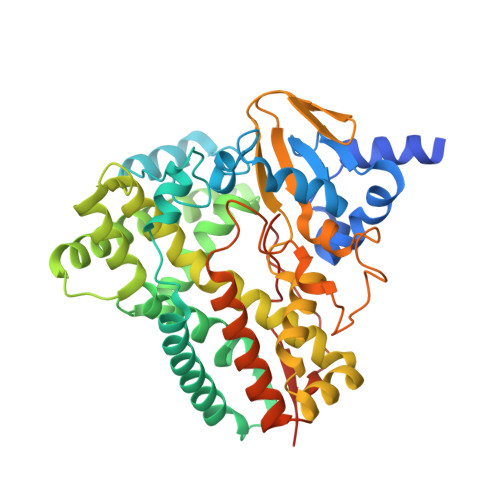
    6RO8

  • PubMed Abstract: 

    Class VII cytochromes P450 are self-sufficient enzymes carrying a phthalate family oxygenase-like reductase domain and a P450 domain fused in a single polypeptide chain. The biocatalytic applications of CYP116B members are limited by the need of the NADPH cofactor and the lack of crystal structures as a starting point for protein engineering. Nevertheless, we demonstrated that the heme domain of CYP116B5 can use hydrogen peroxide as electron donor bypassing the need of NADPH. Here, we report the crystal structure of CYP116B5 heme domain in complex with histidine at 2.6 Å of resolution. The structure reveals the typical P450 fold and a closed conformation with an active site cavity of 284 Å 3 in volume, accommodating a histidine molecule forming a hydrogen bond with the water molecule present as 6th heme iron ligand. MD simulations in the absence of any ligand revealed the opening of a tunnel connecting the active site to the protein surface through the movement of F-, G- and H-helices. A structural alignment with bacterial cytochromes P450 allowed the identification of amino acids in the proximal heme site potentially involved in peroxygenase activity. The availability of the crystal structure provides the bases for the structure-guided design of new biocatalysts.


  • Organizational Affiliation

    Department of Life Sciences and Systems Biology, University of Torino, Via Accademia Albertina 13, Torino, Italy.


Macromolecules
Find similar proteins by:  (by identity cutoff)  |  3D Structure
Entity ID: 1
MoleculeChains Sequence LengthOrganismDetailsImage
Cytochrome P450 RhF449Acinetobacter radioresistensMutation(s): 0 
UniProt
Find proteins for G9BWN9 (Acinetobacter radioresistens)
Explore G9BWN9 
Go to UniProtKB:  G9BWN9
Entity Groups  
Sequence Clusters30% Identity50% Identity70% Identity90% Identity95% Identity100% Identity
UniProt GroupG9BWN9
Sequence Annotations
Expand
  • Reference Sequence
Small Molecules
Ligands 2 Unique
IDChains Name / Formula / InChI Key2D Diagram3D Interactions
HEM
Query on HEM

Download Ideal Coordinates CCD File 
B [auth A]PROTOPORPHYRIN IX CONTAINING FE
C34 H32 Fe N4 O4
KABFMIBPWCXCRK-RGGAHWMASA-L
HIS
Query on HIS

Download Ideal Coordinates CCD File 
C [auth A]HISTIDINE
C6 H10 N3 O2
HNDVDQJCIGZPNO-YFKPBYRVSA-O
Experimental Data & Validation

Experimental Data

  • Method: X-RAY DIFFRACTION
  • Resolution: 2.60 Å
  • R-Value Free: 0.240 
  • R-Value Work: 0.200 
  • Space Group: P 41 21 2
Unit Cell:
Length ( Å )Angle ( ˚ )
a = 109.9α = 90
b = 109.9β = 90
c = 164.2γ = 90
Software Package:
Software NamePurpose
PHENIXrefinement
Cootmodel building
DIALSdata reduction
Aimlessdata scaling
PHASERphasing

Structure Validation

View Full Validation Report


Ligand Structure Quality Assessment 


Entry History 

Deposition Data

Revision History  (Full details and data files)

  • Version 1.0: 2019-08-28
    Type: Initial release
  • Version 1.1: 2019-09-11
    Changes: Data collection, Database references
  • Version 1.2: 2024-01-24
    Changes: Data collection, Database references, Refinement description