6RNS

Crystal structure of the dimerization domain of Gemin5 at 2.7 A


Experimental Data Snapshot

  • Method: X-RAY DIFFRACTION
  • Resolution: 2.69 Å
  • R-Value Free: 0.265 
  • R-Value Work: 0.234 
  • R-Value Observed: 0.235 

wwPDB Validation   3D Report Full Report


This is version 1.2 of the entry. See complete history


Literature

Structural basis for the dimerization of Gemin5 and its role in protein recruitment and translation control.

Moreno-Morcillo, M.Francisco-Velilla, R.Embarc-Buh, A.Fernandez-Chamorro, J.Ramon-Maiques, S.Martinez-Salas, E.

(2020) Nucleic Acids Res 48: 788-801

  • DOI: https://doi.org/10.1093/nar/gkz1126
  • Primary Citation of Related Structures:  
    6RNQ, 6RNS

  • PubMed Abstract: 

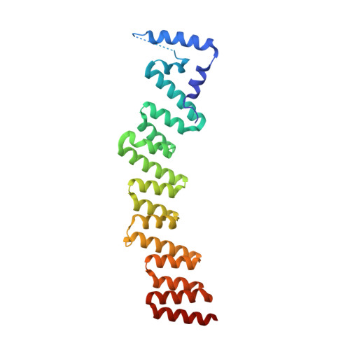
    In all organisms, a selected type of proteins accomplishes critical roles in cellular processes that govern gene expression. The multifunctional protein Gemin5 cooperates in translation control and ribosome binding, besides acting as the RNA-binding protein of the survival of motor neuron (SMN) complex. While these functions reside on distinct domains located at each end of the protein, the structure and function of the middle region remained unknown. Here, we solved the crystal structure of an extended tetratricopeptide (TPR)-like domain in human Gemin5 that self-assembles into a previously unknown canoe-shaped dimer. We further show that the dimerization module is functional in living cells driving the interaction between the viral-induced cleavage fragment p85 and the full-length Gemin5, which anchors splicing and translation members. Disruption of the dimerization surface by a point mutation in the TPR-like domain prevents this interaction and also abrogates translation enhancement induced by p85. The characterization of this unanticipated dimerization domain provides the structural basis for a role of the middle region of Gemin5 as a central hub for protein-protein interactions.


  • Organizational Affiliation

    Centro de Biología Molecular Severo Ochoa, CSIC-UAM, Nicolás Cabrera 1, 28049 Madrid, Spain.


Macromolecules
Find similar proteins by:  (by identity cutoff)  |  3D Structure
Entity ID: 1
MoleculeChains Sequence LengthOrganismDetailsImage
Gem-associated protein 5
A, B
252Homo sapiensMutation(s): 0 
Gene Names: GEMIN5
UniProt & NIH Common Fund Data Resources
Find proteins for Q8TEQ6 (Homo sapiens)
Explore Q8TEQ6 
Go to UniProtKB:  Q8TEQ6
PHAROS:  Q8TEQ6
GTEx:  ENSG00000082516 
Entity Groups  
Sequence Clusters30% Identity50% Identity70% Identity90% Identity95% Identity100% Identity
UniProt GroupQ8TEQ6
Sequence Annotations
Expand
  • Reference Sequence
Small Molecules
Ligands 1 Unique
IDChains Name / Formula / InChI Key2D Diagram3D Interactions
IOD
Query on IOD

Download Ideal Coordinates CCD File 
C [auth A]
D [auth A]
E [auth A]
F [auth A]
G [auth A]
C [auth A],
D [auth A],
E [auth A],
F [auth A],
G [auth A],
H [auth B],
I [auth B],
J [auth B],
K [auth B],
L [auth B],
M [auth B],
N [auth B],
O [auth B]
IODIDE ION
I
XMBWDFGMSWQBCA-UHFFFAOYSA-M
Experimental Data & Validation

Experimental Data

  • Method: X-RAY DIFFRACTION
  • Resolution: 2.69 Å
  • R-Value Free: 0.265 
  • R-Value Work: 0.234 
  • R-Value Observed: 0.235 
  • Space Group: P 1 21 1
Unit Cell:
Length ( Å )Angle ( ˚ )
a = 67.851α = 90
b = 54.622β = 108.818
c = 103.032γ = 90
Software Package:
Software NamePurpose
PHENIXrefinement
AutoSolphasing
XDSdata reduction
Aimlessdata scaling
AutoPROCdata scaling
PHENIXmodel building
Cootmodel building

Structure Validation

View Full Validation Report


Entry History & Funding Information

Deposition Data


Funding OrganizationLocationGrant Number
Spanish Ministry of Science, Innovation, and UniversitiesSpainBFU-2016-80570-R; AEI/FEDER, UE

Revision History  (Full details and data files)

  • Version 1.0: 2019-11-27
    Type: Initial release
  • Version 1.1: 2019-12-18
    Changes: Database references
  • Version 1.2: 2020-01-22
    Changes: Database references