6RA0

A ubiquitin-like dimerization domain controls protein kinase D activation by trans-autophosphorylation


Experimental Data Snapshot

  • Method: X-RAY DIFFRACTION
  • Resolution: 2.26 Å
  • R-Value Free: 0.276 
  • R-Value Work: 0.232 
  • R-Value Observed: 0.234 

wwPDB Validation   3D Report Full Report


This is version 1.2 of the entry. See complete history


Literature

A ubiquitin-like domain controls protein kinase D dimerization and activation by trans-autophosphorylation.

Elsner, D.J.Siess, K.M.Gossenreiter, T.Hartl, M.Leonard, T.A.

(2019) J Biol Chem 294: 14422-14441

  • DOI: https://doi.org/10.1074/jbc.RA119.008713
  • Primary Citation of Related Structures:  
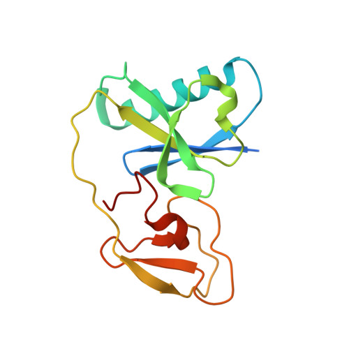
    6RA0

  • PubMed Abstract: 

    Protein kinase D (PKD) is an essential Ser/Thr kinase in animals and controls a variety of diverse cellular functions, including vesicle trafficking and mitogenesis. PKD is activated by recruitment to membranes containing the lipid second messenger diacylglycerol (DAG) and subsequent phosphorylation of its activation loop. Here, we report the crystal structure of the PKD N terminus at 2.2 Å resolution containing a previously unannotated ubiquitin-like domain (ULD), which serves as a dimerization domain. A single point mutation in the dimerization interface of the ULD not only abrogated dimerization in cells but also prevented PKD activation loop phosphorylation upon DAG production. We further show that the kinase domain of PKD dimerizes in a concentration-dependent manner and autophosphorylates on a single residue in its activation loop. We also provide evidence that PKD is expressed at concentrations 2 orders of magnitude below the ULD dissociation constant in mammalian cells. We therefore propose a new model for PKD activation in which the production of DAG leads to the local accumulation of PKD at the membrane, which drives ULD-mediated dimerization and subsequent trans-autophosphorylation of the kinase domain.


  • Organizational Affiliation

    Department of Structural and Computational Biology, Max Perutz Labs, Campus Vienna Biocenter 5, 1030 Vienna, Austria.


Macromolecules
Find similar proteins by:  (by identity cutoff)  |  3D Structure
Entity ID: 1
MoleculeChains Sequence LengthOrganismDetailsImage
Serine/threonine-protein kinase dkf-1153Caenorhabditis elegansMutation(s): 0 
Gene Names: dkf-1W09C5.5
EC: 2.7.11.13
UniProt
Find proteins for Q9XUJ7 (Caenorhabditis elegans)
Explore Q9XUJ7 
Go to UniProtKB:  Q9XUJ7
Entity Groups  
Sequence Clusters30% Identity50% Identity70% Identity90% Identity95% Identity100% Identity
UniProt GroupQ9XUJ7
Sequence Annotations
Expand
  • Reference Sequence
Experimental Data & Validation

Experimental Data

  • Method: X-RAY DIFFRACTION
  • Resolution: 2.26 Å
  • R-Value Free: 0.276 
  • R-Value Work: 0.232 
  • R-Value Observed: 0.234 
  • Space Group: P 21 21 2
Unit Cell:
Length ( Å )Angle ( ˚ )
a = 45.413α = 90
b = 83.018β = 90
c = 37.08γ = 90
Software Package:
Software NamePurpose
PHENIXrefinement
XDSdata reduction
Aimlessdata scaling
PHASERphasing

Structure Validation

View Full Validation Report


Entry History & Funding Information

Deposition Data


Funding OrganizationLocationGrant Number
Austrian Science FundAustriaP28135
Austrian Science FundAustriaP30584

Revision History  (Full details and data files)

  • Version 1.0: 2019-08-14
    Type: Initial release
  • Version 1.1: 2019-08-28
    Changes: Data collection, Database references
  • Version 1.2: 2019-10-09
    Changes: Data collection, Database references