6R6B

Structure of the core Shigella flexneri type III secretion system export gate complex SctRST (Spa24/Spa9/Spa29).


Experimental Data Snapshot

  • Method: ELECTRON MICROSCOPY
  • Resolution: 3.50 Å
  • Aggregation State: PARTICLE 
  • Reconstruction Method: SINGLE PARTICLE 

wwPDB Validation   3D Report Full Report


This is version 1.2 of the entry. See complete history


Literature

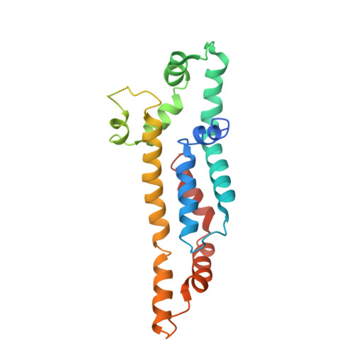
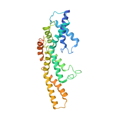
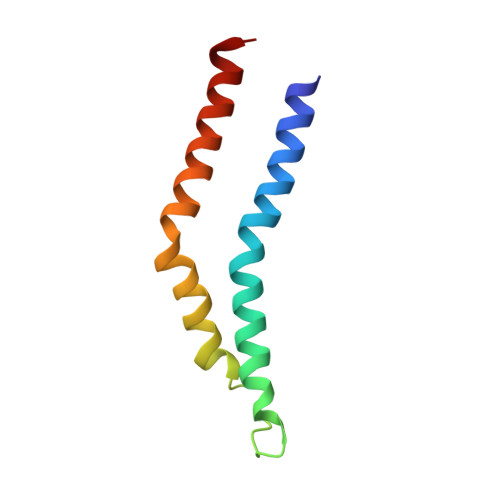
The Structure of an Injectisome Export Gate Demonstrates Conservation of Architecture in the Core Export Gate between Flagellar and Virulence Type III Secretion Systems.

Johnson, S.Kuhlen, L.Deme, J.C.Abrusci, P.Lea, S.M.

(2019) mBio 10

  • DOI: https://doi.org/10.1128/mBio.00818-19
  • Primary Citation of Related Structures:  
    6R69, 6R6B

  • PubMed Abstract: 

    Export of proteins through type III secretion systems (T3SS) is critical for motility and virulence of many major bacterial pathogens. Proteins are exported through a genetically defined export gate complex consisting of three proteins. We have recently shown at 4.2 Å that the flagellar complex of these three putative membrane proteins (FliPQR in flagellar systems, SctRST in virulence systems) assembles into an extramembrane helical assembly that likely seeds correct assembly of the rod. Here we present the structure of an equivalent complex from the Shigella virulence system at 3.5 Å by cryo-electron microscopy. This higher-resolution structure yields a more precise description of the structure and confirms the prediction of structural conservation in this core complex. Analysis of particle heterogeneity also suggests how the SctS/FliQ subunits sequentially assemble in the complex. IMPORTANCE Although predicted on the basis of sequence conservation, the work presented here formally demonstrates that all classes of type III secretion systems, flagellar or virulence, share the same architecture at the level of the core structures. This absolute conservation of the unusual extramembrane structure of the core export gate complex now allows work to move to focusing on both mechanistic studies of type III but also on fundamental studies of how such a complex is assembled.


  • Organizational Affiliation

    Sir William Dunn School of Pathology, University of Oxford, Oxford, United Kingdom.


Macromolecules
Find similar proteins by:  (by identity cutoff)  |  3D Structure
Entity ID: 1
MoleculeChains Sequence LengthOrganismDetailsImage
Surface presentation of antigens protein SpaP
A, B, C, D, E
216Shigella flexneriMutation(s): 0 
Gene Names: spaPspa24CP0153
Membrane Entity: Yes 
UniProt
Find proteins for P0A1L3 (Shigella flexneri)
Explore P0A1L3 
Go to UniProtKB:  P0A1L3
Entity Groups  
Sequence Clusters30% Identity50% Identity70% Identity90% Identity95% Identity100% Identity
UniProt GroupP0A1L3
Sequence Annotations
Expand
  • Reference Sequence
Find similar proteins by:  (by identity cutoff)  |  3D Structure
Entity ID: 2
MoleculeChains Sequence LengthOrganismDetailsImage
Surface presentation of antigens protein SpaR295Shigella flexneriMutation(s): 0 
Gene Names: spaRspa29CP0155
Membrane Entity: Yes 
UniProt
Find proteins for P0A1M6 (Shigella flexneri)
Explore P0A1M6 
Go to UniProtKB:  P0A1M6
Entity Groups  
Sequence Clusters30% Identity50% Identity70% Identity90% Identity95% Identity100% Identity
UniProt GroupP0A1M6
Sequence Annotations
Expand
  • Reference Sequence
Find similar proteins by:  (by identity cutoff)  |  3D Structure
Entity ID: 3
MoleculeChains Sequence LengthOrganismDetailsImage
Surface presentation of antigens protein SpaQG [auth I],
H [auth G],
I [auth H],
J
86Shigella flexneriMutation(s): 0 
Gene Names: spaQspa9CP0154
Membrane Entity: Yes 
UniProt
Find proteins for P0A1M4 (Shigella flexneri)
Explore P0A1M4 
Go to UniProtKB:  P0A1M4
Entity Groups  
Sequence Clusters30% Identity50% Identity70% Identity90% Identity95% Identity100% Identity
UniProt GroupP0A1M4
Sequence Annotations
Expand
  • Reference Sequence
Experimental Data & Validation

Experimental Data

  • Method: ELECTRON MICROSCOPY
  • Resolution: 3.50 Å
  • Aggregation State: PARTICLE 
  • Reconstruction Method: SINGLE PARTICLE 
EM Software:
TaskSoftware PackageVersion
RECONSTRUCTIONRELION3.0
MODEL REFINEMENTPHENIX

Structure Validation

View Full Validation Report


Entry History & Funding Information

Deposition Data


Funding OrganizationLocationGrant Number
Medical Research Council (United Kingdom)United KingdomM011984
Wellcome TrustUnited Kingdom201536
Wellcome TrustUnited Kingdom109136
Wellcome TrustUnited Kingdom201536
Wolfson FoundationUnited KingdomWL160052

Revision History  (Full details and data files)

  • Version 1.0: 2019-05-29
    Type: Initial release
  • Version 1.1: 2019-07-10
    Changes: Data collection, Database references
  • Version 1.2: 2019-12-18
    Changes: Other