6QZO

Crystal structure of DyP-type peroxidase from Cellulomonas bogoriensis


Experimental Data Snapshot

  • Method: X-RAY DIFFRACTION
  • Resolution: 2.40 Å
  • R-Value Free: 0.267 
  • R-Value Work: 0.242 
  • R-Value Observed: 0.243 

wwPDB Validation   3D Report Full Report


This is version 1.2 of the entry. See complete history


Literature

Characterization of a New DyP-Peroxidase from the Alkaliphilic Cellulomonad, Cellulomonas bogoriensis.

Habib, M.H.Rozeboom, H.J.Fraaije, M.W.

(2019) Molecules 24

  • DOI: https://doi.org/10.3390/molecules24071208
  • Primary Citation of Related Structures:  
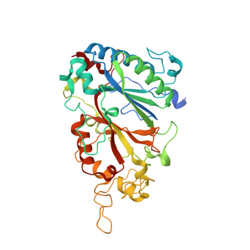
    6QZO

  • PubMed Abstract: 

    DyP-type peroxidases are heme-containing enzymes that have received increasing attention over recent years with regards to their potential as biocatalysts. A novel DyP-type peroxidase ( Cbo DyP) was discovered from the alkaliphilic cellulomonad, Cellulomonas bogoriensis , which could be overexpressed in Escherichia coli . The biochemical characterization of the recombinant enzyme showed that it is a heme-containing enzyme capable to act as a peroxidase on several dyes. With the tested substrates, the enzyme is most active at acidic pH values and is quite tolerant towards solvents. The crystal structure of Cbo DyP was solved which revealed atomic details of the dimeric heme-containing enzyme. A peculiar feature of Cbo DyP is the presence of a glutamate in the active site which in most other DyPs is an aspartate, being part of the DyP-typifying sequence motif GXXDG. The E201D Cbo DyP mutant was prepared and analyzed which revealed that the mutant enzyme shows a significantly higher activity on several dyes when compared with the wild-type enzyme.


  • Organizational Affiliation

    Molecular Enzymology, Groningen Biomolecular Sciences and Biotechnology Institute, University of Groningen, Nijenborgh 4, 9747AG Groningen, The Netherlands. m.habib@rug.nl.


Macromolecules
Find similar proteins by:  (by identity cutoff)  |  3D Structure
Entity ID: 1
MoleculeChains Sequence LengthOrganismDetailsImage
Peroxidase
A, B, C, D, E
A, B, C, D, E, F, G, H
383Cellulomonas bogoriensis 69B4 = DSM 16987Mutation(s): 0 
Gene Names: N869_13850
UniProt
Find proteins for A0A0A0BZU2 (Cellulomonas bogoriensis 69B4 = DSM 16987)
Explore A0A0A0BZU2 
Go to UniProtKB:  A0A0A0BZU2
Entity Groups  
Sequence Clusters30% Identity50% Identity70% Identity90% Identity95% Identity100% Identity
UniProt GroupA0A0A0BZU2
Sequence Annotations
Expand
  • Reference Sequence
Small Molecules
Ligands 2 Unique
IDChains Name / Formula / InChI Key2D Diagram3D Interactions
HEM
Query on HEM

Download Ideal Coordinates CCD File 
I [auth A]
J [auth B]
K [auth C]
L [auth D]
M [auth E]
I [auth A],
J [auth B],
K [auth C],
L [auth D],
M [auth E],
N [auth F],
O [auth G],
P [auth H]
PROTOPORPHYRIN IX CONTAINING FE
C34 H32 Fe N4 O4
KABFMIBPWCXCRK-RGGAHWMASA-L
PGE
Query on PGE

Download Ideal Coordinates CCD File 
Q [auth H]TRIETHYLENE GLYCOL
C6 H14 O4
ZIBGPFATKBEMQZ-UHFFFAOYSA-N
Experimental Data & Validation

Experimental Data

  • Method: X-RAY DIFFRACTION
  • Resolution: 2.40 Å
  • R-Value Free: 0.267 
  • R-Value Work: 0.242 
  • R-Value Observed: 0.243 
  • Space Group: P 62
Unit Cell:
Length ( Å )Angle ( ˚ )
a = 173.989α = 90
b = 173.989β = 90
c = 283.003γ = 120
Software Package:
Software NamePurpose
REFMACrefinement
iMOSFLMdata reduction
Aimlessdata scaling
PHASERphasing

Structure Validation

View Full Validation Report


Entry History 

Deposition Data

Revision History  (Full details and data files)

  • Version 1.0: 2019-04-03
    Type: Initial release
  • Version 1.1: 2019-04-10
    Changes: Data collection, Database references
  • Version 1.2: 2024-01-24
    Changes: Data collection, Database references, Refinement description