6QWN

Protein peptide complex


Experimental Data Snapshot

  • Method: X-RAY DIFFRACTION
  • Resolution: 3.89 Å
  • R-Value Free: 0.329 
  • R-Value Work: 0.282 
  • R-Value Observed: 0.284 

wwPDB Validation   3D Report Full Report


Ligand Structure Quality Assessment 


This is version 2.0 of the entry. See complete history


Literature

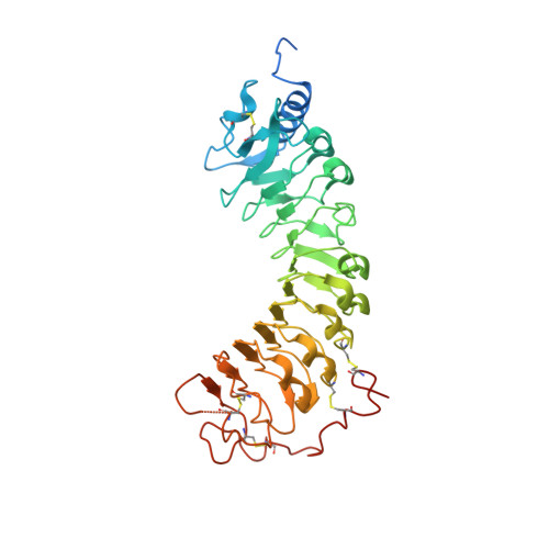
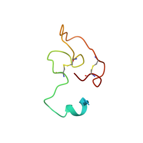
Structural basis for recognition of RALF peptides by LRX proteins during pollen tube growth.

Moussu, S.Broyart, C.Santos-Fernandez, G.Augustin, S.Wehrle, S.Grossniklaus, U.Santiago, J.

(2020) Proc Natl Acad Sci U S A 117: 7494-7503

  • DOI: https://doi.org/10.1073/pnas.2000100117
  • Primary Citation of Related Structures:  
    6QWN, 6QXP, 6TME

  • PubMed Abstract: 

    Plant reproduction relies on the highly regulated growth of the pollen tube for sperm delivery. This process is controlled by secreted RALF signaling peptides, which have previously been shown to be perceived by Catharanthus roseus RLK1-like ( Cr RLK1Ls) membrane receptor-kinases/LORELEI-like GLYCOLPHOSPHATIDYLINOSITOL (GPI)-ANCHORED PROTEINS (LLG) complexes, or by leucine-rich repeat (LRR) extensin proteins (LRXs). Here, we demonstrate that RALF peptides fold into bioactive, disulfide bond-stabilized proteins that bind the LRR domain of LRX proteins with low nanomolar affinity. Crystal structures of LRX2-RALF4 and LRX8-RALF4 complexes at 3.2- and 3.9-Å resolution, respectively, reveal a dimeric arrangement of LRX proteins, with each monomer binding one folded RALF peptide. Structure-based mutations targeting the LRX-RALF4 complex interface, or the RALF4 fold, reduce RALF4 binding to LRX8 in vitro and RALF4 function in growing pollen tubes. Mutants targeting the disulfide-bond stabilized LRX dimer interface fail to rescue lrx infertility phenotypes. Quantitative biochemical assays reveal that RALF4 binds LLGs and LRX cell-wall modules with drastically different binding affinities, and with distinct and mutually exclusive binding modes. Our biochemical, structural, and genetic analyses reveal a complex signaling network by which RALF ligands instruct different signaling proteins using distinct targeting mechanisms.


  • Organizational Affiliation

    The Plant Signaling Mechanisms Laboratory, Department of Plant Molecular Biology, University of Lausanne, 1015 Lausanne, Switzerland.


Macromolecules
Find similar proteins by:  (by identity cutoff)  |  3D Structure
Entity ID: 1
MoleculeChains Sequence LengthOrganismDetailsImage
Pollen-specific leucine-rich repeat extensin-like protein 1A,
C [auth B],
E [auth C],
G [auth D],
I [auth E]
379Arabidopsis thalianaMutation(s): 0 
Gene Names: PEX1At3g19020K13E13.23
UniProt
Find proteins for Q9LJ64 (Arabidopsis thaliana)
Explore Q9LJ64 
Go to UniProtKB:  Q9LJ64
Entity Groups  
Sequence Clusters30% Identity50% Identity70% Identity90% Identity95% Identity100% Identity
UniProt GroupQ9LJ64
Sequence Annotations
Expand
  • Reference Sequence
Find similar proteins by:  (by identity cutoff)  |  3D Structure
Entity ID: 2
MoleculeChains Sequence LengthOrganismDetailsImage
Protein RALF-like 4B [auth F],
D [auth G],
F [auth H],
H [auth I],
J
56Arabidopsis thalianaMutation(s): 0 
Gene Names: RALFL4At1g28270F3H9.8
UniProt
Find proteins for Q9FZA0 (Arabidopsis thaliana)
Explore Q9FZA0 
Go to UniProtKB:  Q9FZA0
Entity Groups  
Sequence Clusters30% Identity50% Identity70% Identity90% Identity95% Identity100% Identity
UniProt GroupQ9FZA0
Sequence Annotations
Expand
  • Reference Sequence
Oligosaccharides

Help

Entity ID: 3
MoleculeChains Length2D Diagram Glycosylation3D Interactions
alpha-D-mannopyranose-(1-3)-beta-D-mannopyranose-(1-4)-2-acetamido-2-deoxy-beta-D-glucopyranose-(1-4)-2-acetamido-2-deoxy-beta-D-glucopyranose
K
4N-Glycosylation
Glycosylation Resources
GlyTouCan:  G81315DD
GlyCosmos:  G81315DD
GlyGen:  G81315DD
Entity ID: 4
MoleculeChains Length2D Diagram Glycosylation3D Interactions
2-acetamido-2-deoxy-beta-D-glucopyranose-(1-3)-2-acetamido-2-deoxy-beta-D-glucopyranose
L
2N-Glycosylation
Glycosylation Resources
GlyTouCan:  G79924RM
GlyCosmos:  G79924RM
GlyGen:  G79924RM
Entity ID: 5
MoleculeChains Length2D Diagram Glycosylation3D Interactions
beta-D-mannopyranose-(1-4)-2-acetamido-2-deoxy-beta-D-glucopyranose-(1-6)-2-acetamido-2-deoxy-beta-D-glucopyranose
M
3N-Glycosylation
Glycosylation Resources
GlyTouCan:  G62916OK
GlyCosmos:  G62916OK
GlyGen:  G62916OK
Entity ID: 6
MoleculeChains Length2D Diagram Glycosylation3D Interactions
2-acetamido-2-deoxy-beta-D-glucopyranose-(1-4)-2-acetamido-2-deoxy-beta-D-glucopyranose
N
2N-Glycosylation
Glycosylation Resources
GlyTouCan:  G42666HT
GlyCosmos:  G42666HT
GlyGen:  G42666HT
Entity ID: 7
MoleculeChains Length2D Diagram Glycosylation3D Interactions
beta-D-mannopyranose-(1-4)-2-acetamido-2-deoxy-beta-D-glucopyranose-(1-4)-2-acetamido-2-deoxy-beta-D-glucopyranose
O
3N-Glycosylation
Glycosylation Resources
GlyTouCan:  G15407YE
GlyCosmos:  G15407YE
GlyGen:  G15407YE
Experimental Data & Validation

Experimental Data

  • Method: X-RAY DIFFRACTION
  • Resolution: 3.89 Å
  • R-Value Free: 0.329 
  • R-Value Work: 0.282 
  • R-Value Observed: 0.284 
  • Space Group: C 1 2 1
Unit Cell:
Length ( Å )Angle ( ˚ )
a = 205.841α = 90
b = 114.091β = 116.25
c = 146.977γ = 90
Software Package:
Software NamePurpose
PHENIXrefinement
XDSdata reduction
XDSdata scaling
PHASERphasing

Structure Validation

View Full Validation Report


Ligand Structure Quality Assessment 


Entry History & Funding Information

Deposition Data


Funding OrganizationLocationGrant Number
European Research CouncilSwitzerland716358

Revision History  (Full details and data files)

  • Version 1.0: 2020-03-18
    Type: Initial release
  • Version 1.1: 2020-03-25
    Changes: Database references
  • Version 1.2: 2020-04-08
    Changes: Database references
  • Version 2.0: 2020-07-29
    Type: Remediation
    Reason: Carbohydrate remediation
    Changes: Atomic model, Data collection, Derived calculations, Structure summary